Download
1 / 20
360 likes | 1.16k Views
Fluorescence Resonance Energy Transfer ( FRET). Xingwei Wang. FRET based immunosensor. From ref [1]. Principle. Two fluorophores: Donor & acceptor In close proximity the donor absorbs energy from the source transfers the energy to the acceptor the acceptor emits fluorescent energy

E N D
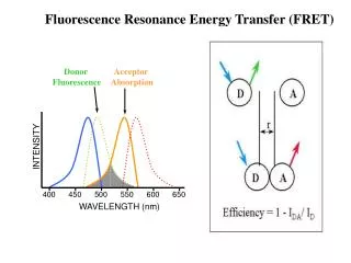
Fluorescence Resonance Energy Transfer (FRET) Xingwei Wang
FRET based immunosensor From ref [1]
Principle • Two fluorophores: Donor & acceptor • In close proximity • the donor absorbs energy from the source • transfers the energy to the acceptor • the acceptor emits fluorescent energy • Distance dependent property • Detect conformational changes when antibodies combine with their respective antigens
Principle (2) • The fluorophores were conjugated to an antibody-Protein A complex • then immobilized to the distal end of an optical fiber. • Conformational changes • Investigate donor and acceptor fluorophore emission spectrum
Application I: Monitor early markers of myocardial infarction • 1.1 million cases of acute myocardial infarction (AMI) occur each year in the United States • Can be modified and inserted subcutaneously to provide early warning of an impending heart attack
Principle • Försters distance: the distance where energy transfer from the donor to acceptor fluorophore is 50% (< 100 A) • Close: λ0 -> λ2 • Separated: λ0 -> λ1 • Conformational Change
Performance • Detection limit: 27nM • 600 µm diameter silica core optical fibers • Taper end: • hydrofluoric acid for 2-4 hours • 12.0 mm of the cladding was removed • Evanescent wave reaches the sensing area of the cladding-stripped fiber tip • Exciting the donor fluorophores located within its penetrating depth
Emission Spectrum From ref [1]
Spectrum Methods • The donor fluorophore excitation light: 540 nm • Peak 1 (P1), is the donor emission spectrum with maximum peak intensity at 570 nm. • Peak 2 (P2), is the acceptor emission spectrum with maximum peak intensity at 610 nm. • Rather than analyzing intensity of the emission curves • susceptible to instrumental baseline shifts • Using • the maximum area under each emission spectrum • The ratio of the maximum donor to acceptor area (P1/P2)
Results • A decrease in the P1/P2 ratio after antigen addition is indicative of energy transfer.
Problem - High STD • Different tapering angles - different amounts of photons being captured back • Different exposed surface areas - different antibody-Protein immobilized – different signal strength
Applications II: Food safety: Detection of Listeria • U.S. each year • 33 million cases of foodborne diseases • more than 5 billion dollars for treatment • about 9,000 deaths • Listeria - one of the main organisms causing the outbreaks of foodborne illnesses • Rapid, accurate methods for detecting pathogens in food processing facilities are needed.
Advantage • Detect only viable analytes • Reduce false positives • Listeria antigen detection limits: 2.0µg/ml
Spectrum • I(h = 570 nm to 575 nm): the average fluorescence intensity of the donor fluorophore • I(h =608 nm to 613 nm): the average fluorescence intensity of the acceptor
Measuremet With no antigen present (baseline) With specific or nonspecific antigen present Ratio used to determine change
Advantages • Portable • On-site analysis of samples • Reduce the large economical burden by food products recalls and medical treatments
FRET video • http://www.youtube.com/watch?v=pMH8zcWa7WA
References • Development of a FRET based fiber-optic biosensor for early detection of myocardial infarctionPierce, M.E.; Grant, S.A.;Engineering in Medicine and Biology Society, 2004. EMBC 2004. Conference Proceedings. 26th Annual International Conference of theVolume 1, 2004 Page(s):2098 - 2101 Vol.3 • Development of a novel FRET immunosensor for detection of listeriaKo, S.; Grant, S.A.;Sensors, 2003. Proceedings of IEEEVolume 1, 22-24 Oct. 2003 Page(s):288 - 292 Vol.