Download
1 / 138
1.44k likes | 1.8k Views
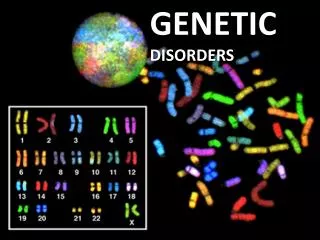
Genetic disorders. Dr.K.V.Bharathi. Normal karyotype. Study of chromosomes karyotyping A karyotype is the standard arrangement of a photographed, stained chromosomes pairs which are arranged in order of decreasing length. When chromosomes are preparing to divide, the DNA

E N D
Genetic disorders Dr.K.V.Bharathi
Normal karyotype • Study of chromosomeskaryotyping • A karyotype is the standard arrangement of a photographed, stained chromosomes pairs which are arranged in order of decreasing length
When chromosomes are preparing to divide, the DNA replicates itself into two strands called chromatids Replicating chromosome The same chromosome under normal conditions Telomere Centromere The two chromatids Telomere
Chromosome nomenclature • Two arms • p (petite) small and q (follows p in alphabet) • 1-22 = autosome numbers • X, Y = sex chromosomes
Cytogenetic terminology • Short arm p and long arm q • Each Chromosome is divided into 2 or more regions • Each region is subdivided into bands and sub-bands • Total no of chromosomes is given first followed by sex chromosome and finally description of abnormality in ascending order.eg:47,XY,+21 ,and Xp 21.2 • 46,XY,del(16)(p11.2 p13.1)
The normal human karyotype • Somatic cells: 22 pairs of autososmes & 1 pair of sex chromosomes (46,XX or 46,XY). • The normal karyotype is diploid (2 copies of each chromosome). • Sperm & eggs carry 23 chromosomes & are haploid (one copy of each chromosome).
What is the difference between an Autosome and a Sex-chromosome? • Autosomes are the first 22 homologous pairs of human chromosomes that do not influence the sex of an individual. • Sex Chromosomes are the 23rd pair of chromosomes that determine the sex of an individual.
Sperm determines genotypic sex by contributing either an X or a Y chromosome during fertilization.
46,XX = female 46,XY = male Giemsa banding (G-banding)
Three classes of chromosome • Metacentric - centromere in middle • Submetacentric - centromere distant from middle • Acrocentric - centromere at end
Uses of karyotype analysis: • Genotypic sex ( identification of X & Y chromosomes). • Ploidy ( euploid, aneuploid or polyploid). • Chromosomal structural defects (translocation, isochromosome, deletion etc..).
Some definitions • Haploid (n)- refers to a single set of chromosomes (23 in humans).Sperm & eggs are haploid. • Diploid (2n)- refers to a double set of chromosomes (46 in humans). Somatic cells are diploid. • Euploid- refers to any multiple of the haploid set of chromosomes (from n-8n)
Polyploid- refers to any multiple of the haploid set of chromosomes> diploid (2n). • Aneuploid- refers to karyotypes that do not have multiples of the haploid set of chromosomes. • Monosomy- refers to an aneuploid karyotype with one missing chromosome (XO in Turner’s syndrome). • Trisomy- refers to an aneuploid karyotype with one extra chromosome (trisomy 21 in Down’s syndrome))
Aneuploidy results from the failure of chromosomes to separate normally during cell division: Meiotic Nondisjunction
4N First meiotic division NORMAL SEPARATION 2N Second meiotic division N Gametes Fertilization 2N Zygotes NORMAL ZYGOTE
First meiotic division NONDISJUNCTION Second meiotic division Gametes Fertilization Zygotes TRISOMIC ZYGOTE MONOSOMIC ZYGOTE
Aneuploidy usually results from non-disjunction • Chromosomes or chromatids fails to separate • An error of mitotic or meiotic spindle attachment to centromere • May occur in either the maternal or the paternal germ cells • More commonly arises in the mother • Frequency of non-disjunction increases with maternal age
Six main types • Deletion • Ring chromosome • Duplication • Isochromosome • Inversion • paracentric & pericentric • Translocation • Robertsonian & reciprocal
Deletion • Involves loss of part of a chromosome • Results in monosomy of that chromosomal segment • Clinical effects due to • Insufficient gene products • Unmasking of mutant alleles on normal chromosome Before deletion After deletion
Two types of deletion Terminal Interstitial
Ring chromosomeBreaks occur in both arms of a chromosome. The two broken ends anneal; the two acentric fragments are lost. Results in double deletion (in p and in q).
Isochromosome Mirror image chromosome Loss of one arm with duplication of other Loss of p-arm Duplication of q-arm
Inversion Two breaks in one chromosome The fragment generated rotates 180o and reinserts into the chromosome Pericentric - involves p and q arm Paracentric - involves only one arm
Translocation - exchange of chromosomal material between two or more chromosomes • Reciprocal • Robertsonian • If no essential chromosome material lost or genes damaged then the individual is clinically normal • However, there is an increased chance of chromosomally unbalanced offspring
Reciprocal Translocation • Involves two chromosomes • One break in each chromosome • The two chromosomes exchangebroken segments Before translocation After translocation
Robertsonian translocation • Named after W. R. B. Robertson who first identified them in grasshoppers in 1916 • Most common structural chromosome abnormality in humans • Frequency = 1/1000 livebirths • Involves two acrocentric chromosomes • Two types • Homologous acrocentrics involved • Non-Homologous acrocentrics involved
Homologous acrocentric, i.e. chromosome 14 lost + = Non-homologous acrocentric, i.e. chromosomes 14 & 21 lost + =
A balanced chromosome 14 & 21 Robertsonian translocation
What is mutation? • A mutation may be defined as a permanent change in the DNA. • These structural DNA changes affect protein expression & function.
Mutations affect protein synthesis Transcription:Mutated DNA will produce faulty mRNA leading to the production of a faulty protein.
Somatic & Germ cell mutations Mutations that occur in somatic cells such as skin cells or hair are termed Somatic. Germline mutations occur only in the gametes. These mutations are more threatening because they can be passed to offspring .
Germline mutations can be transmitted to future generations. • Those that occur in somatic cells may contribute to the pathogenesis of neoplasia. • Drugs, chemical & physical agents that increase the rate of mutation act as carcinogens.
Mutagens are agents that cause mutations. They include: 1. High Temperatures 2. Toxic Chemicals (pesticides, etc) 3. Radiation (nuclear and solar)
Types of mutations Chromosomal mutation: affecting whole or a part of a chromosome Gene mutation: changes to the bases in the DNA of one gene
Major types of genetic mutations • Point mutations: Single base substitutions . • Frameshift mutations: base pair insertions or deletions that change the codon reading frame. • Large deletions: can result in loss of gene or juxtapose genes to create a hybrid that encodes a new “fusion” protein. • Expansion of trinucleotide repeats:can arise in genes that have repeated sequences. Affected patients can have 100s or 1000s of repeats (normal:10-30).
Gene Mutations: DNA base alterations Point mutation- eg:sickle cell anemia Insertion Deletion Inversion Frame Shifts
Point mutation - when a base is replaced with a different base. CGG CCC AAT to CGG CGC AATGuanine for Cytosine Insertion - when a base is added CGG CCC AAT to CGG CGC CAA TGuanine is added Deletion - the loss of a base CGG CCC AAT to CGG CCA A T loss of Cytosine
Frame Shift mutations • A frame shift mutation results from a base deletion or insertion. Each of these changes the triplets that follow the mutation. CGG CCC AAT to CGG CGC CAA T • Frame shift mutations have greater effects than a point mutation because they involve more triplets. • This in turn changes the amino acids of the protein!
Classification of genetic disorders 1.Gross chromosomal abnormalities 2.Diseases with multifactorial inheritance 3.Disorders related to mutant genes of large effect
Trisomy 21 - Down's Syndrome - karyotype 47, XX +21 or 47, XY+21 - frequency about 1 in 600 births
Trisomy 18 - Edward's Syndrome • - karyotype 47, XX +18 or 47, XY+18 • - frequency about 1 in 8,000births
Trisomy 13 - Patau's Syndrome • - karyotype 47, XX +13 or 47, XY+13 • - frequency about 1 in 10,000 births
Sex chromosome trisomies • - 47, XXY (Klinefelter Syndrome), 47,XXX, 47,XYY • Triploidies of other chromosomes • Rare • usually incompatible with life