Download
1 / 8
80 likes | 170 Views
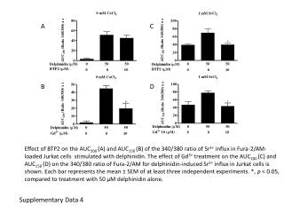
Appendix Supplementary data (online only) to: Marleen Kok, Wilbert Zwart, Caroline Holm, Renske Fles, Michael Hauptmann, Laura J. Van ’t Veer, Lodewyk F.A. Wessels, Jacques Neefjes, Olle Stål, Sabine C. Linn, Göran Landberg, Rob Michalides

E N D
Appendix Supplementary data (online only) to: Marleen Kok, Wilbert Zwart, Caroline Holm, Renske Fles, Michael Hauptmann, Laura J. Van ’t Veer, Lodewyk F.A. Wessels, Jacques Neefjes, Olle Stål, Sabine C. Linn, Göran Landberg, Rob Michalides PKA-induced phosphorylation of ERα at serine 305 and/or high PAK1 levels predict tamoxifen resistance in the majority of ER positive breast cancer cases
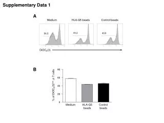
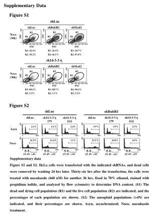
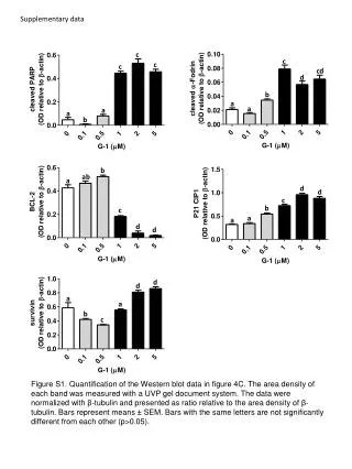
Figure A1. Staining for the catalytic subunit of PKA phosphorylated at threonine 197. A) Representative example of positive pPKA staining. B) Absence of pPKA expression after dephosphorylation via incubation with lambda phosphatase on the tumor slide prior to immunohistochemistry. A B
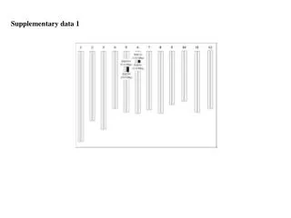
Figure A2. Flow diagram of patients Flow of patients through the study including number of patients in each stage. Reasons for dropout and number of events (recurrence) in each subgroup are listed. Start of trial 564 randomized patients 262 events 70 excluded (no FFPE tissue and no data on ERα) 32 events 494 with ERα data 230 events 110 excluded (ERα ≤10%) 55 events 384 ERα >10% 175 events 153 excluded (no ERα305-P, pPKA and PAK1 data) 71 events 231 with ERα305-P, pPKA or PAK1 data 104 events
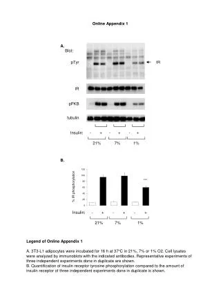
Figure A3. Overall tamoxifen benefit in patients included in the current translational study (n=231) compared to tamoxifen benefit in all ER-positive breast cancer patients included in the trial (n=384). Kaplan-Meier analysis of recurrence-free survival according to randomization in A) 384 patients of whom tumor material was available and the ER was expressed in >10% of tumor cells and in B) 231 patients of whom tumor material was available, the ER was expressed in >10% of tumor cells and data on PAK1, PKA and ER305-P were available. ---- tamoxifen, --- no adjuvant systemic treatment N=231 Log-rank=0.020 HR=0.63, 95% CI 0.42-0.93, p=0.021 N=384 Log-rank=0.003 HR=0.64, 95% CI 0.47-0.86, p=0.004
Table A2. Distribution of pPKA according to clinicopathological parameters (training series NKI)
Table A3 . Randomized controlled trial (Lund) used as validation series. Distribution of clinico-pathological parameters in the excluded cases (ERα>10%, (no data to generate PAK1-PKA/ ERS305-P Score, n=153) vs included patients in the validation series (n=231). * According Nottingham Grading system (Elston et al.).
Table A4. Distribution of prognostic factors in the subgroup of tumors classified as sensitive by the PAK1-PKA/ ERS305-P Score in the validation set (PAK1 and/or PKA/ER305-P negative) according to systemic treatment received (tamoxifen versus no adjuvant systemic treatment). * According Nottingham Grading system (Elston et al.). † Mann-Whitney U test ‡ Fisher’s Exact test Abbreviations: LN lymph node status, ER estrogen receptor, PR progesterone receptor, IDC invasive ductal carcinoma, ILC invasive lobular carcinoma.