Download
1 / 48
480 likes | 556 Views
Explore the cardiovascular system, heart anatomy, blood flow pathways, and heart surgery procedures. Learn about heart attacks and different aspects of cardiac health. Discover the importance of valves and the significance of the cardiac cycle.

E N D
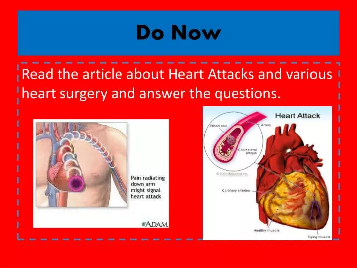
Do Now Read the article about Heart Attacks and various heart surgery and answer the questions.
Cardiovascular System Ch.13 The Veinest of the Systems
Objectives • Identify the organs of the cardiovascular system and it’s overall function. • Label the different parts of the heart. • Describe the pathway of blood.
The Cardiovascular System • Pumps 7,000 liters of a blood a DAY!!! • Functions to carry oxygen and nutrients to the cells of the body and carry carbon dioxide and other waste away from the cells of the body.
Structures • Heart • pumps blood to the lungs and throughout the body • Arteries, arterioles • Caries blood AWAY from the heart to cells • Capillaries • Place where nutrient and gas waste exchanges • Veins, venules • Carries blood TO the heart from cells
Arteries vs. Veins • Arteries • THICK! • Veins • Thinner • Contain 1-way valves.
Pulmonary vs. Systemic Circuits • Pulmonary • Carries Oxygen depleted blood to the lungs to pick up O2 and unload CO2. • Systemic • Sends oxygen-rich blood from heart to all body cells.
The Heart • Located in the thoracic cavity, rests on top of the diaphragm • Encased in a “parietal pericardium”
Wall of the Heart 1. Outer Epicardium • Made of epithelial tissue • Protects the heart by reducing friction 2. Middle Myocardium • Mostly cardiac tissue involved in pumping blood out of the heart 3. Inner Endocardium • Made of epithelial tissue • Lines the inner chambers of the heart
Heart Chambers • Atria • Upper chambers • Receive blood returning to the heart • Ventricles • Lower chambers • Receive blood from atria and force blood into arteries • Septum • Separates left and right so blood doesn’t mix
Heart Valves • Atrioventricular Valves: separate atria from ventricles and prevent back-flow of blood. • Tricuspid: • Right side • Bicuspid/Mitral: • Left side
Mitral Valve Prolapse • Heart “murmur” • Mitral valve contracts and stretches bulging into the left atrium • Blood regurgitates back into the atrium • Normal Heart Beat • Mitral regurgitation
Heart Valves • Semilunar Valves: • Pulmonary: • Found between the right ventricle and the pulmonary artery • Aortic: • Found between the left ventricle and the aortic arch
Do Now Write 3 things you know about the eye or structure of the eye. Quiz is postponed until tomorrow (Tuesday).
Objectives • Explain the flow of blood through the heart, lungs, and body. • Compare and contrast angina vs. a heart attack.
The Heart • Take out your heart diagram so we can finish labeling!
Pathway of Blood • Follow along on your worksheet! • Great Pathway of Blood Video!
Angina Pectoris • A “thrombus” or “embolus” blocks or narrows a coronary artery and deprives myocardial cells of Oxygen, causing pain. • *many people mistake this for a heart attack. • A complete blockage by a blood clot is a myocardial infarction (aka heart attack)
Do Now • Hand in your anatomy valentine if you haven’t done so already! • Clean all purses/bags/books off of the desks so you don’t get them dirty! • Take a copy of the “heart practical study guide” from my desk as well as a lab dissection packet.
Objectives • Identify the structures of the sheep heart. • Trace the path of blood flow through a sheep heart. • Explain the importance of the valves. • Understand what will be on the lab practical.
Sheep Heart Dissection! • HAPPY VALENTINE’S DAY! Today we will be dissecting a sheep heart. • Be sure to FOLLOW ALONG with the lab and go through each and every structure that it tells you to look at, you will be tested on them on both the hearts and the heart models. • You may use your phones to take pictures • Practical will be on Tuesday!
Do Now • What part of the heart pumps oxygenated blood to the body and what “tube” does it go through? • Name 2 differences between arteries and veins. • What is another name for the visceral pericardium?
Objectives • To explain the flow of blood through both pulmonary and systemic circuits. • To understand the cardiac cycle. • To explain what causes your “heart beat”.
Blood Flow • SmartNotebook File
Do Now • WITHOUT TALKING…… put yourselves in order of the flow of blood through the body up at the front of the room.
Do Now • What do you think causes your heart beat? • Write the flow of blood ONLY through the heart.
Objectives • To explain what occurs in a normal cardiac cycle. • To define systole and diastole. • To measure ones pulse to determine their heart rate. • To determine what causes heart sounds.
Cardiac Cycle • Systole= contraction • Diastole = relaxing * During atrial systole, ventricles are in ventricular diastole * During ventricular systole, atria are in atrial diastole • Both relax briefly after ventricular systole • Cycle Animation
Steps of the Cycle • Pressure is low during ventricular diastole, opening the A-V valves (tricuspid/bicuspid) • Ventricles fill with blood • A-V valves close when ventricular pressure exceeds atrial pressure • Papillary muscles pull on chordae tendinae to prevent valves from bulging back into atria (which would cause a murmur) • During ventricular systole, atrial pressure is low and they begin to fill up again
Steps of the Cycle 5) Ventricular pressure rises, opening the semilunar valves, forcing blood into the pulmonary trunk and aortic arch 6) Pressure drops in ventricles after contraction, and the semilunar valves close. Echocardiogram
Heart Sounds • The heart makes a “Lubb-Dupp” sound • The “Lubb” is the closing of the A-V valve during ventricular contraction. • The “Dupp” is the closing of the semilunar valve when the ventricles are relaxing.
Pulse of Life Lab • You will be measuring your pulse and seeing how your heart rate is affected by different activities! • You will turn in the series of questions upon completing the lab as well as construct a graph.
Do Now • Explain the steps of the cardiac cycle. • How do you think these steps are coordinated?
Objectives • To explain the cardiac conduction system • To identify the components of an EKG • To identify different heart arrhythmias
Cardiac Muscle Fibers • Cardiac muscle fibers form a network called a functional synctium which contracts as a unit.
Cardiac Conduction System - Coordinates the events of the cardiac cycle
Cardiac Conduction System • The Synotrial Node (SA Node)- “Pacemaker” • Specialized cardiac muscle tissue • Can reach threshold on its own • Generates impulses 70-80 times per minute • Atrial synctium- causes atrial contraction • A-V Node • A-V Bundle (Bundle of His) • Purkinje Fibers- causes ventricular contraction
Cardiac Conduction • Conduction
Electrocardiogram • Recording of the electrical changes that occur in the myocardium (cardiac muscle)
Electrocardiogram • P-wave: depolarization of atria • QRS: depolarization of ventricle fibers (R=Left, S=right) • T-wave: repolarization of ventricles
Researching Arrhythmias • V-fib (Ventricular Fibrillation) • Sinus Bradycardia • Sinus Tachycardia • Atrial Fibrillation • Atrial Flutter • Asystole
Do Now • In your own words, Explain the cardiac conduction system and how it works to control the cardiac cycle.
Objectives • To identify EKG’s of different heart arrhythmias. • To determine how fast a heart is beating by looking at an EKG. • To explain blood pressure readings and understand how blood pressure can be effected.
Reading an EKG • Read the article about reading an EKG! • How can you figure out the rate?
Blood Pressure (13.5) • Blood pressure= the force blood exerts against the inner walls of blood vessels. • Interesting Fact! The human heart creates enough pressure to squirt blood 30 feet!!
Measuring Blood Pressure • Systolic Pressure: max pressure during ventricular systole • Diastolic Pressure: max pressure during ventricular diastole
What gives us a “pulse”? • Ventricular contraction causes a “surge” in arteries, distending the elastic arterial walls, pressure drops immediately after contraction • Only felt in arteries close to the surface such as your carotid in your neck.
Factors Affecting Blood Pressure • Heart Action • Stroke volume (vol of blood discharged with each contraction) and heart rate • Blood Volume • If you lose a lot of blood, your blood pressure will be lower. • Peripheral Resistance • If the vessels are constricted, pressure increased • Blood Viscosity • More viscous=more pressure