Download

1 / 39

E N D
OBJECTIVES - To study the anatomy of articular structures - To study about the ligaments strengthening it - To study the major relations of knee joint - To study the blood supply of knee joint - To study the nerve supply of knee joint - To study the applied aspects of knee joint
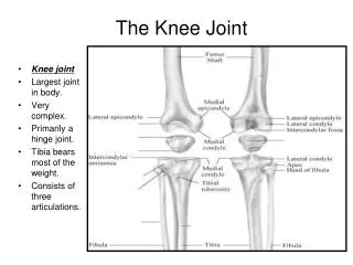
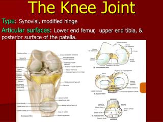
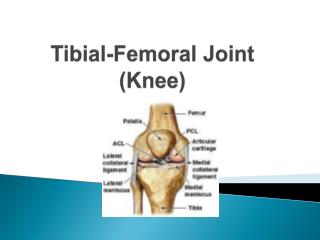
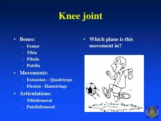
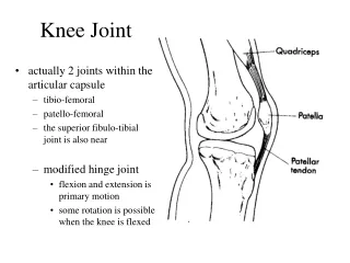
KNEE JOINT Type- - It is a compound synovial joint Articular surface- - The condyles of femur - The condyles of tibia - The patella Largest and more complex joint of the body
LIGAMENTS 1) Fibrous capsule- - is very thin and deficient anteriorly - posteriorly it is attached to the Intercondylar line - attached to the intercondylar ridge of tibia - capsule present between menisci and tibia is called as coronary ligament
LIGAMENTS 2) Ligamentum patellae- - central portion of the common tendon of quadriceps femoris - remaining portion forms medial and lateral retinacula - ligament is about 7.5cm long and 2.5cm broad - attached to the apex of the patella - below to the upper part of the tibialtuberosity - superficial and deep infrapatellar bursae related to it
LIGAMENTS 3) Tibial collateral ligament(medial ligament)- - long band of great strength - superiorly attached to the medial epicondyle - inferiorly to the medial border and shaft of tibia - superficial part is about 10cm long and 1.25cm broad - posterior part blends with the capsule and meniscus
LIGAMENTS 4)Fibular collateral ligament(lateral ligament)- - ligament is strong and cord like - it is about 5cm long - superiorly attached to the lateral epicondyle of femur - inferiorly to the head of the fibula - separated from lateral meniscus by tendon of popliteus
LIGAMENTS 5) Oblique popliteal ligament- - is an expansion from the tendon of semimembranosus - runs upwards and posteriorly, blends with the capsule - attached to intercondylar line and lateral condyle 6) Arcuate popliteal ligament- - posterior expansion of short lateral ligament - extends backward from the head of fibula - arches over tendon of popliteus - attached to intercondylar area of tibia
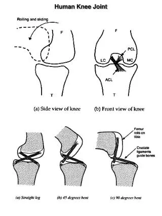
LIGAMENTS 7) Anterior cruciate ligaments- - this are very thick and strong fibrous band - direct bonds of union between tibia and femur - begins from anterior part of intercondylar area of tibia - runs upwards, backwards and laterally - attached to posterior part of medial surface of lateral condyle
LIGAMENTS 8) Posterior cruciate ligaments- - begins from the posterior part of intercondylar area - runs upward, forward and medially - attached to the ant: part of lateral surface of medial condyle of femur - both ACL and PCL maintain stability of knee joint
LIGAMENTS 9) Menisci(semilunar cartilages)- - They are two fibrocartilaginous discs. - they are shaped like crescents - they deepen the articular surface of tibia - has two ends attached to the tibia - two borders, outer border is thick and convex fixed to capsule - inner border is thin, concave and free
LIGAMENTS Menisci- - two surfaces, upper surface is concave - the lower surface is flat - the peripheral thick part is vascular - inner part is avascular and is nourished by synovial fluid - medial meniscus is nearly semicircular - posterior fibres of anterior end are continuous with transverse ligament - lateral meniscus is nearly circular, posterior end attached to the femur through meniscofemoral ligament
LIGAMENTS • Function of menisci- - because of their flexibility they can adapt their contour to the varying curvature - the menisci serve as shock absorbers - they help in lubricating the joint cavity - nerve supply result in sensory function too
LIGAMENTS 11) Transverse ligaments- - connects the anterior ends of medial and lateral menisci • Synovial membrane- - it lines the capsule, except posteriorly - forming a common covering for cruciate ligaments - in front it is absent from the patella - above patella it forms suprapatellar bursa - below patella it covers infrapatellar pad of fat
BURSAE Anterior- - subcutaneous prepatellar bursa - subcutaneous infrapatellar bursa - deep infrapatellar bursa - suprapatellar bursa Lateral- - deep to lateral head of gastrocnemius - between fibular collateral ligament and biceps femoris
BURSAE - Between fibular collateral ligament and popliteus - between popliteus and lateral condyle of femur Medial- - deep to the medial head of gastrocnemius - anserine bursa separates Sartorius, Gracilis, semitentinosus from the tibia and tibial collateral ligament - deep to tibial collateral ligament - deep to semimembranosus
RELATIONS Anteriorly- - anterior bursae - ligamentum patellae - patellar plexus of nerves Posteriorly- - popliteal vessels - tibial nerve - middle genicular vessels and nerves
RELATIONS Posterolaterally- - lateral head of gastrocnemius - plantaris - common peroneal nerve Posteromedially- - medial head of gastrocnemius - semitendinosus - semimembranosus, gracilis, popliteus
RELATIONS Medially- - Sartorius - great saphenous vein - saphenous nerve - medial genicular vessels and nerve Laterally- - biceps femoris - lateral genicular vessels and nerves
BLOOD SUPPLY • The knee joint is supplied by the anastomoses around it - genicular branches of popliteal artery - descending genicular branch of femoral artery - descending branch of lateral circumflex femoral artery - recurrent branches of anterior tibial artery - circumflex fibular branch of posterior tibial artery
NERVE SUPPLY - Femoral nerve - sciatic nerve - obturator nerve
CLINICAL ANATOMY • Stability is maintained by - cruciate ligament maintains anteroposterior stability - collateral ligament maintains side to side stability - iliotibial tract also stabilises the knee joint - fibrous capsule • Genu valgum- abnormal abduction due to abnormality in angle between thigh and leg • Genu varum- abnormal adduction of knee
CLINICAL ANATOMY • Osteoarthritis of knee joint • medial meniscus is more vulnerable to injury • anterior cruciate ligament is more commonly damaged
SUMMARY - It is the weight bearing joint of lower limb - Deformities of the joint affects the stability - It is a compound synovial joint - Osteoarthritis of knee joint is common during old age