Download

1 / 19
210 likes | 619 Views
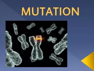
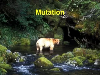
Mutation. Mutations. Any change in the DNA sequence of an organism is a mutation. Mutations are the source of the altered versions of genes that provide the raw material for evolution.

E N D
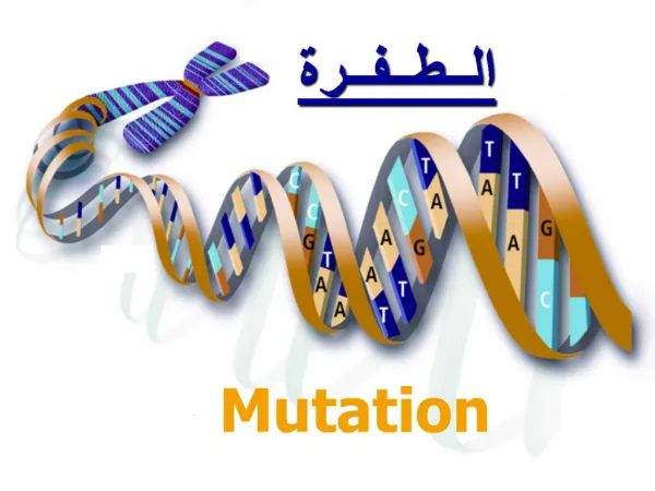
Mutations • Any change in the DNA sequence of an organism is a mutation. • Mutations are the source of the altered versions of genes that provide the raw material for evolution. • Most mutations have no effect on the organism, especially among the eukaryotes, because a large portion of the DNA is not in genes and thus does not affect the organism’s phenotype. • Of the mutations that do affect the phenotype, the most common effect of mutations is lethality, because most genes are necessary for life. • Only a small percentage of mutations causes a visible but non-lethal change in the phenotype.
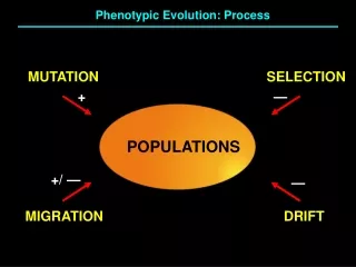
Mutations are Random • A central tenet of biology is that the flow of information from DNA to protein is one way. DNA cannot be altered in a directed way by changing the environment. Only random DNA changes occur. • The “fluctuation test”, an early experiment in bacterial genetics (Luria and Delbruck, 1943) showed that variations in bacterial phenotypes are due to pre-existing mutations and not due to physiological changes induced by environmental conditions. • A large batch of E. coli is infected with phage T1. Most are lysed by the phage, but a few survive. Are the survivors a few rare individuals who managed to induce their T1-protection mechanisms on time, or are they pre-existing mutants? • --if protection is a physiological condition induced by the presence of the phage, then the percentage of survivors will be the same among all small batches of the cells; all cells are genetically identical. • if protection is due to pre-exisiting mutations, then some small batches will have many survivors (descendants of T1-resistant mutants), while most other batches will have few or no survivors (there were no T1-resistant mutants in the original batch). • Result: some batches had many survivors, but most batches had few or none. T1-resistant mutants existed in the population before T1 was added.
Types of DNA Change • The simplest mutations are base changes, where one base is converted to another. These can be classified as either: • --“transitions”, where one purine is changed to another purine (A -> G, for example), or one pyrimidine is changed to another pyrimidine (T -> C, for example). • “transversions”, where a purine is substituted for a pyrimidine, or a pyrimidine is substituted for a purine. For example, A -> C. • Another simple type of mutation is the gain or loss f one or a few bases. • Larger mutations include insertion of whole new sequences, often due to movements of transposable elements in the DNA or to chromosome changes such as inversions or translocations. • Deletions of large segments of DNA also occurs.
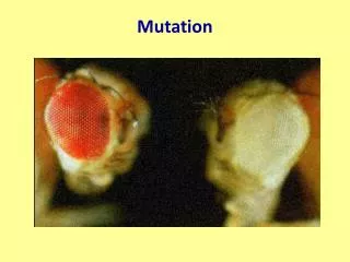
Types of Mutation • Mutations can be classified according to their effects on the protein (or mRNA) produced by the gene that is mutated. • 1. silent mutations (synonymous mutations). Since the genetic code is degenerate, several codons produce the same amino acid. Especially, third base changes often have no effect on the amino acid sequence of the protein. These mutations affect the DNA but not the protein. Therefore they have no effect on the organism’s phenotype. • 2. missense mutations. Missense mutations substitute one amino acid for another. Some missense mutations have very large effects, while others have minimal or no effect. It depends on where the mutation occurs in the protein’s structure, and how big a change in the type of amino acid it is. • Example: HbS, sickle cell hemoglobin, is a change in the beta-globin gene, where a GAG codon is converted to GUG. GAG codes for glutamic acid, which is a hydrophilic amino acid that carries a -1 charge, and GUG codes for valine, a hydrophobic amino acid. This amino acid is on the surface of the globin molecule, exposed to water. Under low oxygen conditions, valine’s affinity for hydrophobic environments causes the hemoglobin to crystallize out of solution.
More Types of Mutation • 3. Nonsense mutations convert an amino acid into a stop codon. The effect is to shorten the resulting protein. Sometimes this has only a little effect, as the ends of proteins are often relatively unimportant to function. However, often nonsense mutations result in completely non-functional proteins. • an example: Hb-β McKees Rock. Normal beta-globin is 146 amino acids long. In this mutation, codon 145 UAU (codes for tyrosine) is mutated to UAA (stop). The final protein is thus 143 amino acids long. The clinical effect is to cause overproduction of red blood cells, resulting in thick blood subject to abnormal clotting and bleeding. • 4. Sense mutations are the opposite of nonsense mutations. Here, a stop codon is converted into an amino acid codon. Since DNA outside of protein-coding regions contains an average of 3 stop codons per 64, the translation process usually stops after producing a slightly longer protein. • Example: Hb-α Constant Spring. alpha-globin is normally 141 amino acids long. In this mutation, the stop codon UAA is converted to CAA (glutamine). The resulting protein gains 31 additional amino acids before it reaches the next stop codon. This results in thalassemia, a severe form of anemia.
Frameshifts and Reversions • Translation occurs codon by codon, examining nucleotides in groups of 3. If a nucleotide or two is added or removed, the groupings of the codons is altered. This is a “frameshift” mutation, where the reading frame of the ribosome is altered. • Frameshift mutations result in all amino acids downstream from the mutation site being completely different from wild type. These proteins are generally non-functional. • example Hb-α Wayne. The final codons of the alpha globin chain are usually AAA UAC CGU UAA, which code for lysine-tyrosine-arginine-stop. In the mutant, one of the A’s in the first codon is deleted, resulting in altered codons: AAU ACC GUU AAG, for asparagine-threonine-valine-lysine. There are also 5 more new amino acids added to this, until the next stop codon is reached. • A “reversion” is a second mutation that reverse the effects of an initial mutation, bringing the phenotype back to wild type (or almost). • Frameshift mutations sometimes have “second site reversions”, where a second frameshift downstream from the first frameshift reverses the effect. • Example: consider Hb Wayne above. If another mutation occurred that added a G between the 2 C’s in the second codon, the resulting codons would be: AAU ACG CGU UAA, or asparagine-threonine-arginine-stop. Note that the last 2 codons are back to the original. Two amino acids are still altered, but the main mutational effect has been reverted to wild type.
mRNA Problems • Although many mutations affect the protein sequence directly, it is possible to affect the protein without altering the codons. • Splicing mutations. Intron removal requires several specific sequences. Most importantly, introns are expected to start with GT and end in AG. Several beta globin mutations alter one of these bases. The result is that one of the 2 introns is not spliced out of the mRNA. The polypeptide translated from these mRNAs is very different from normal globin, resulting in severe anemia. • Polyadenylation site mutations. The primary RNA transcript of a gene is cleaved at the poly-A addition site, and 100-200 A’s are added to the 3’ end of the RNA. If this site is altered, an abnormally long and unstable mRNA results. Several beta globin mutations alter this site: one example is AATAAA -> AACAAA. Moderate anemia was the result.
Trinucleotide Repeats • A fairly new type of mutation has been described, in which a particular codon is repeated. • During replication, DNA polymerase can “stutter” when it replicates several tandem copies of a short sequence. For example, CAGCAGCAGCAG, 4 copies of CAG, will occasionally be converted to 3 copies or 5 copies by DNA polymerase stuttering. • Outside of genes, this effect produces useful genetic markers called SSR (simple sequence repeats). • Within a gene, this effect can cause certain amino acids to be repeated many times within the protein. In some cases this causes disease • For example, Huntington’s disease is a neurological disease that generally strikes in middle age, producing paranoia, uncontrolled limb movements, psychosis, and death. Woody Guthrie, a folk singer from the 1930’s, had this disease. • The Huntington’s disease gene normally has between 11 and 33 copies of CAG (codon for glutamine) in a row. The number occasionally changes. People with HD have 37 or more copies, up to 200). The rate of copy number change is much higher in HD people--too many copies makes the repeated sequence more subject to DNA polymerase stuttering during meiosis. • Interestingly, the age of onset of the disease is related to the number of CAG repeats present: the more repeats, the earlier the onset.
Germinal vs. Somatic Mutations • Back up to the phenotype level • Mutations can occur in any cell. They only affect future generations if they occur in the cells that produce the gametes: these are “germinal” or “germ line” mutations. • Mutations in other cells are rarely noticed, except in the case of cancer, where the mutated cell proliferates uncontrollably. Mutations in cells other than germ line cells are “somatic” mutations. • A human body contains 1013 - 1014 cells approximately. The average mutation rate for any given nucleotide is about 1 in 109. That is, on the average 1 cell in 109 has that particular nucleotide altered. This means that virtually every possible base change mutation occurs repeatedly in our body cells.
Retinoblastoma • Retinoblastoma is a hereditary form of cancer that illustrates the interaction between somatic and germinal mutations. • This disease affects the retinoblasts, cells that are precursors to the retinal cells. Retinoblasts exist in the eyes until about 3 years of age. Thus, retinoblastoma always occurs by about this age. • There are 2 forms of retinoblastoma: the hereditary form, which is almost always bilateral (affects both eyes), and the spontaneous form, which almost always affects just one eye. Neither parent has the disease in spontaneous cases. • Why should the hereditary form affect both eyes while the spontaneous form affects only 1 eye?
RB Explanation • The retinoblastoma gene, Rb, is a “tumor suppressor” gene. Individual cells become cancerous if they lack this gene, or if both copies are defective mutants: Rb- Rb-. • The mutation rate for the Rb gene is about 10-6, which means that about 1 copy in 106 will spontaneously go from Rb+ to Rb-. • The retina contains about 108 retinoblasts. • So, since we are diploid, a cell must go from Rb+ Rb+ to Rb+ Rb- and then to Rb- Rb- to become cancerous. This requires 2 independent mutations. Since the chance of 2 independent events is the product of the individual chances, the chance of a Rb+ Rb+ cell becoming Rb- Rb- is 10-6 * 10-6 = 10-12. Given that there are 108 retinoblasts per person, this would occur about 1 time in 10,000 people. This is about the rate of occurrence of spontaneous RB. That is, about 1 person in 10,000 will have 1 cell that is homozygous mutant, resulting in RB in one eye only.
More RB Explanation • People with hereditary RB inherit one mutant allele. Every cell in their bodies starts out Rb+ Rb-. It takes only a single somatic mutation to convert a cell to Rb- Rb-. • Given that the mutation rate is about 10-6 and the number of cells per retina is about 108, it is almost a certainty that multiple tumors will start in both eyes of people with hereditary retinblastoma.
Determining the Human Mutation Rate • Not easy. Need to look at dominant or co-dominant mutations, because humans can’t be test-crossed. • One study in Michigan looked for dominant mutations known to be caused by a single gene, with no known phenocopies that are fully expressed and highly penetrant. They looked at achondroplasia (dwarfism) and retinoblastoma. • Achondroplasia: found 7 new (non-hereditary) cases among 242,257 births, for a rate of 1.4 x 10⁻⁵ new mutations per allele per generation. • Retinoblastoma: found 52 new cases among 1,054,985 births, for a rate of 2.3 x 10⁻⁵ new mutations per allele per generation. • Protein electrophoresis: found 4 new alleles among 1,226,099 examined, for a rate of 3.3 x 10⁻⁶ new mutations per allele per generation. A bit lower than for the dominant mutations, but these genes are smaller.
More on Mutation Rate • In 1945, at the end of World War 2, the US detonated 2 nuclear weapons over Hiroshima and Nagasaki Japan. • Extensive studies were done of the genetic effects on the survivors. A number of genes were examined by looking at the proteins they produced by gel electrophoresis. • Radiation levels: a dose of 1 Sievert (Sv) is equal to 100 rem in the old terminology. • A dose of radiation that would kill 50% of people within 60days is about 5 Sv. • Natural exposure in Chicago are is about 1 milliSievert (1 mSv) per year. • Natural exposure in Denver (5000 foot altitude) is about 1.8 mSv/year. • radiation workers are permitted up to 20 mSv per year. • average Hiroshima survivor: 200 mSv
Hiroshima Study • In the largest biochemical genetics study, 3 new mutations affecting migration rates of proteins on electrophoresis gels were found among 667,404 alleles examined among Hiroshima survivors. Also, 3 new alleles were found among 466,881 alleles examined in the control group. • No easily detected change in mutation rate. Possible to estimate radiation dose needed to double human mutation rate at 4 Sv (with lethal dose of 5 Sv). • However, cancer of all types is increased among survivors and continues high to the present.
Detecting Mutagens • Radiation and certain chemical compounds are “mutagens”: they cause mutation. • Cancer is caused by somatic mutations, and so mutagens are also carcinogens. • Testing for mutagenicity is a key step is development of pharmaceutical drugs. • Simple test using bacteria (Salmonella, a close relative of E. coli) developed by Bruce Ames: the “Ames test”.
Ames Test • Start with Salmonella that are his-, auxotrophs unable to make their own histidine. They will only grow if histidine is added to the growth medium. • Add compound to be tested to growth medium, count number of colonies growing. These are revertants, which have been mutated back to wild type. • In many cases, mutagens need to be activated, converted to mutagenic state, by enzymes in the liver that are meant to detoxify dangerous compounds. Liver extracts are often added to the growth medium to accomplish this. • Test isn’t perfect: Salmonella are prokaryotes, and we have complex biochemistries that modify foreign compounds. But, it is a good initial screen.