1 / 32
320 likes | 324 Views
Students should be able to:<br>- describe the structure of the eye as seen in front view and in horizontal section <br>- state the principal functions of component parts of the eye in producing a focused image of near <br>and distant objects on the retina <br>- describe the pupil reflex in response to bright and dim light

E N D
2 Mechanisms that take place in the eye 1) Pupil reflex (controls the amount of light that enters the eyes) 2) Accommodation (focusing)
1) Pupil reflex - controls the amount of light that enters the eyes
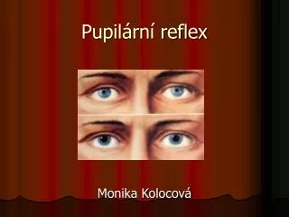
Pupil enlarges when? Controlling light entry into eye Pupil dims when?
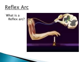
Controlling entry of light into eye ◼ Through altering diameter of the pupil ◼ Size of pupil controlled by 2 sets of involuntary muscles (in iris) ◼ One set arranged in a circle round the pupil ◼ One set arranged radially ◼ Antagonistic → circular contract; radial relax → pupil smaller
How pupil reflex works… Light intensity Pupil Circular muscle Radial muscle Becomes smaller High Contracts Relaxes Becomes bigger Low Relaxes Contracts
2) Accommodation (focusing)
Accommodation Definition: The adjustment of the lens of the eye so that clear images of objects at different distances are formed on the retina
Normal vision occurs when light is focused directly on the retina rather than in front or behind it
Distant object Suspensory ligaments Near object
Focusing for far vision (>7m) - parallel rays - flatter; less convex - circular muscle relaxes
Focusing for near vision - diverging rays - more convex - circular muscle contracts
How accommodation works… Object Light rays Lens Suspensory ligaments Ciliary Muscles Thinner; less convex Thicker; more convex Parallel Distant Taut Relax Contract Slacken Near Diverging
Near point ◼ Object so close to eye ◼ Requires ciliary muscle to contract fully ◼ Suspensory ligaments slacken ◼ Lens becomes most convex ◼ Beyond this point, image is blurred (lens cannot be adjusted anymore)
Stereoscopic vision ◼ Visual field of our 2 eyes overlap ◼ 2 eyes may focus on same object but different image received by each eye ◼ Brain able to interpret images together to obtain a 3D view ◼ Gives us a better perception of size, depth and distance of the object from us
Is seeing believing?
Optical illusion1 Stare at the pattern for 30 seconds or longer. Than immediately look at a blank sheet of white paper.
What do you see? ◼ You should see the same pattern in reverse ◼ Why? ◼ While you are staring, the photoreceptors in your retina get tired—especially the cells viewing the white part of the pattern. When you switch your gaze, the least tired cells take over, producing the brightest part of the afterimage
Optical illusion2 Is it really a square?
Optical illusion3 Are the centre circles equal sizes?
Optical illusion4 Are the red lines of different lengths?
Answers: It is a square The two center circles are exactly the same size The two red lines are exactly the same length Go ahead. Use a ruler to make sure So what's going on? In each case your brain is comparing the item in question with the surrounding patterns—and misinterpreting the information. Perception is not always reality.
Resources ◼ Anatomy of the eye http://www.discover-the-eye.com/dae060305en.swf ◼ http://www.physpharm.fmd.uwo.ca/undergrad/medsweb/ L1Eye/Eye.swf