Download
1 / 2
20 likes | 22 Views
OptraSCAN offers artificial intelligence & machine learning-based System for accurate, rapid, and reproducible analysis of Prostate Cancer. <br>Contact us at- info@optrascan.com<br>Visit- https://www.optrascan.com/solutions/prostate-cancer-biomarker-analysis

E N D
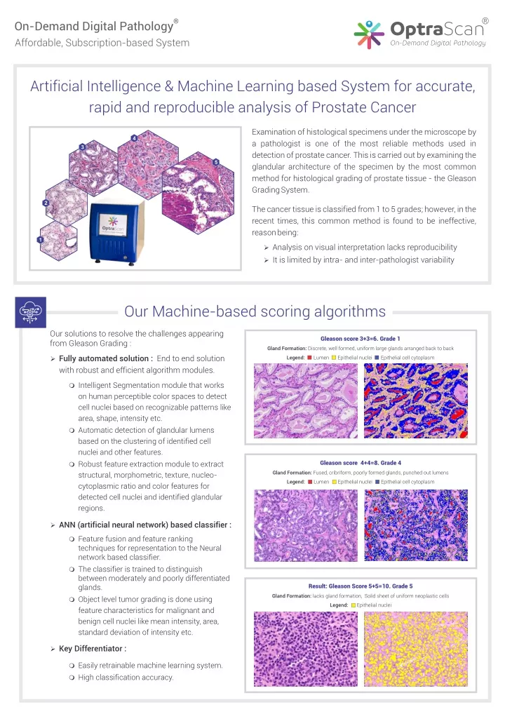
® ® OptraScan On-Demand Digital Pathology Affordable, Subscription-based System Artificial Intelligence & Machine Learning based System for accurate, rapid and reproducible analysis of Prostate Cancer Examination of histological specimens under the microscope by a pathologist is one of the most reliable methods used in detection of prostate cancer. This is carried out by examining the glandular architecture of the specimen by the most common method for histological grading of prostate tissue - the Gleason Grading System. 4 3 5 2 The cancer tissue is classified from 1 to 5 grades; however, in the recent times, this common method is found to be ineffective, reason being: 1 Ø Analysis on visual interpretation lacks reproducibility Ø It is limited by intra- and inter-pathologist variability Our Machine-based scoring algorithms Our solutions to resolve the challenges appearing from Gleason Grading : Gleason score 3+3=6. Grade 1 Gland Formation: Discrete, well formed, uniform large glands arranged back to back Legend: Lumen Epithelial nuclei Epithelial cell cytoplasm Ø Fully automated solution : End to end solution with robust and efficient algorithm modules. m Intelligent Segmentation module that works on human perceptible color spaces to detect cell nuclei based on recognizable patterns like area, shape, intensity etc. m Automatic detection of glandular lumens based on the clustering of identified cell nuclei and other features. m Robust feature extraction module to extract structural, morphometric, texture, nucleo- cytoplasmic ratio and color features for detected cell nuclei and identified glandular regions. Gleason score 4+4=8. Grade 4 Gland Formation: Fused, cribriform, poorly formed glands, punched out lumens Legend: Lumen Epithelial nuclei Epithelial cell cytoplasm Ø ANN (artificial neural network) based classifier : m Feature fusion and feature ranking techniques for representation to the Neural network based classifier. m The classifier is trained to distinguish between moderately and poorly differentiated glands. m Object level tumor grading is done using feature characteristics for malignant and benign cell nuclei like mean intensity, area, standard deviation of intensity etc. Result: Gleason Score 5+5=10. Grade 5 Gland Formation: lacks gland formation, Solid sheet of uniform neoplastic cells Legend: Epithelial nuclei Ø Key Differentiator : m Easily retrainable machine learning system. m High classification accuracy.
® OptraScan ® On-Demand Digital Pathology Solutions OS-15 OS-FS OS-FL OS-120 15-slide brightfield 7-slide frozen sections, with live view mode 15-slide fluorescence, with 6 filter cubes 120-slide brightfield ® IMAGEPath Web-based Image Management and Viewing TM OptraASSAYS TM TELEPath Web and Mobile Digital Conferencing On-Demand Image Analysis ® CLOUDPath Laboratory Information Management System 100 Century Center Court, Suite 410, San Jose, CA 95112 OptraSCAN is an ISO13485 certified company *All OptraSCAN systems and solutions are for research use only