Download
1 / 7
70 likes | 74 Views
One study used OCT to classify fibrous, calcified, and lipid plaques. Fibrous plaques were characterized as having a signal-rich area with low backscattering. Calcium plaques were identified as having a signal-poor area, and lipid plaques were identified as having a signal-rich layer overlying a lipid core. Lipidic and calcified plaques had lower classification accuracy than fibrous plaques. However, these variations were explained by the lack of distinction between these three plaques.

E N D
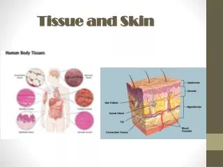
Preparing and Analyzing Oct Tissue scigenscigenus 3 hours ago 6 min read The oct tissue is a biological sample of tissues derived from various organs, including the liver, heart, and lung. It can be used for diagnostic tests and research. Several methods are available for extraction, preparation, and analysis. Some of the most important processes in its preparation and analysis are covered in this article. Preparation
When freezing tissue, using a solid matrix is a good idea. This is especially true for tissue samples that may be used for RNA extraction or protein extraction. Oct tissue Compound provides a convenient specimen matrix and helps eliminate unwanted background staining. The oct tissue compound is a water-soluble blend of glycols and resins. Although the container is small, the compound's speedy freeze-down is a boon to researchers and surgeons. It is available in 4 oz squeeze bottles. ● ● The oct tissue Compound is an excellent choice for embedding fresh or frozen tissue. In addition to its quick and easy freeze-down, the compound's unique formula will prevent dulling of microtome knives and leave no residue on slides during staining. An adequately designed cryomold will provide a good environment for the best possible tissue sample preparation. Make sure to label the container with its contents and orientation, and ensure that it is stored on powdered dry ice. Making a cold isopentane and dry ice bath is the first stage in preparing a frozen sample. It should be poured over the tissue sample and left to sit for 15 to 30 seconds. ● ● ● ● After the bath has been thoroughly cooled, a frozen block should be transferred to the cryotome cryostat. Proper block orientation will ensure that each section is on the same side, and the smallest unit will be on the bottom of the mold. When processing the specimen, many variables affect the quality of the section. For example, the number of cells in the sample will influence the number of units produced. Furthermore, the morphology of the tissues will be altered by prolonged fixation. Fixation
Tissue fixation is a process in which the sample is fixed to preserve the tissue components in a "life-like state." This prevents autolysis and promotes the preservation of morphology, structural features, antigens, and chemical composition. Tissue fixation is a critical step in histopathology. It allows us to study the changes that occur to the tissue after processing. Choosing the correct fixative is essential to the optimal outcome of histopathology. The ideal fixative should preserve the extracellular morphology, cellular morphology, and antigens in a life-like state. In addition to preserving cell morphology, a fixative should not denature proteins or enzymes important for histopathology. When choosing a fixative, it is best to choose a neutral buffer. 10% Neutral Buffered Formalin (NBF) is commonly used for histology. However, 10% NBF can be toxic to the respiratory system and eyes. The typical fixation time is 4 - 24 hours. Fixation for longer than this period may lead to over-fixation. Over-fixation results in loss of signal and can mask the antigen. The use of microwave irradiation for fixing tissue is another option. Several studies have reported the effectiveness of microwave irradiation in fixing tissues. Microwave irradiation of tissue was performed on 140 paired tissue sections from non-neoplastic and neoplastic tissues. Results indicated that microwave irradiation did not cause any loss of erythrocytes. However, the maximum loss of tissues was seen in lymphoid tissues. For long-term tissue preservation, it is preferable to use a paraformaldehyde solution. Paraformaldehyde is also a suitable fixative for special stains.
Fixation should be carried out immediately after surgery. The longer the fixation time, the more background noise and fixation artifacts will be observed. If the fixative is not sufficiently stable, it may not be reproducible. Incubation The incubation of oct tissue is a process that involves numerous steps. It is important to select the correct procedure for each tissue type. This will allow optimal specific binding of antibodies to the target tissues and will minimize background staining. Tissue is typically dissected into one or more high-quality sections, which can then be encased in an oct tissue box. A suitable fixative is added to ensure that the sample is kept intact. As with other types of experiments, there are many variables to be considered. However, performing an appropriate number of microscopic examinations is not a substitute for detailed information. Some of the more complex procedures, such as freezing, may require optimization. For instance, the duration of the freeze-thaw cycle should be measured, and the storage conditions adjusted to optimize the process. The correct volume of the requisite fixative should be determined. Generally, the largest volume of a given solution is required to produce an adequate level of fixation. An example application is the alpha-1 antitrypsin deficiency in the liver. OCT compound is an effective reagent used to stabilize and preserve the tissue during freezing. The most appropriate compound is the best way to ensure optimal preservation of the contents and the best chance of a successful freeze.
This protocol is a bit complex and requires specialized equipment, but the benefits are well worth the investment. Besides the standard freezing, RNA extraction and protein extraction can be performed on samples. In the end, the results provide valuable insights into how the body metabolizes its constituent parts. Detection of secretory mucins, glycolipids, and glycan epitopes A large family of glycoproteins, known as mucins, has been identified in many human epithelial cancers. Mucins are secreted extracellular proteins with anti-adhesive properties. They are often expressed on the surface of cells and are highly glycosylated. It is believed that mucins may have a role in maintaining a barrier and limiting contact with pathogens and antigens. These glycoproteins are composed of O-linked glycans covalently attached to proteins, lipids, or other glycoproteins. Changes in the composition of glycans and the glycosylation pathways are associated with tumorigenesis and contribute to disease development. In particular, the immune system recognizes foreign carbohydrate epitopes and responds by binding and internalizing them. The composition of glycans is altered in parallel with changes in cellular metabolism, and alterations in the structure of glycan structures play an important role in cancer development. For example, tumors display abnormal core fucosylation and N-glycan branching. One type of glycan that has been shown to exhibit aberrantly high expression in adenocarcinomas is MUC1. This glycoprotein is a founding member of the mucin family. The
core of mucins is comprised of a large extracellular domain with multiple repeats. There are also five potential O-glycosylation sites within the domain. Mucins are synthesized in both cis and trans-Golgi compartments. The primary glycan of MUC1 is an N-acetylgalactosamine (GalNAc) in b1,3-linkage with galactose. However, additional sugar structures can be added to the initial glycan. In addition to being a source of anti-adhesive agents, mucins also provide a scaffold for the growth and maintenance of tissue architecture. This is because they can be anchored into the plasma membrane via the posttranslational attachment of lipids. Another potential function of mucins is to facilitate migration. Diagnostic utility Optical coherence tomography (OCT) is a noninvasive, marker-free, light-based optical tool that provides high-resolution cross-sectional images of the tissue. OCT can be used in a wide variety of applications. Some of the main uses of OCT include imaging blood vessels and soft tissue. It can also be used to help identify fungal elements in onychomycosis. While OCT is useful for various conditions, there are some limitations. Most commonly, it is not able to detect pigmented lesions. Moreover, the recorded signal lacks specificity and may change after fixation. This makes it less suitable for use in the diagnosis of tumors. However, oct tissue can be used to assess the degree of fibrosis. Furthermore, it is capable of providing architectural information. Combined with fluorescence, OCT can identify structures not captured by traditional histology.
Advanced techniques have been developed to improve the subcellular level resolution. These advanced techniques can also improve the accuracy of diagnosis by providing tissue-specific contrast. They can provide information such as tissue stiffness and molecular content. Ultimately, they can be combined with computational processing to deliver real-time evaluations. There are many possible applications for OCT in the larynx. Among them, it is possible to use the technology to visualize the epithelial layer of the larynx, to differentiate between pre-malignant changes, and to diagnose cancer. In addition, Oct tissue can be a valuable adjunct during therapeutic interventions in the larynx. Another potential application of Oct tissue in the larynx is its ability to perform a targeted biopsy. This can be achieved using fibreoptic trans-nasal OCT. The operating canal of a flexible laryngoscope can be used to introduce a biocompatible probe.