Download
1 / 89
920 likes | 1.22k Views
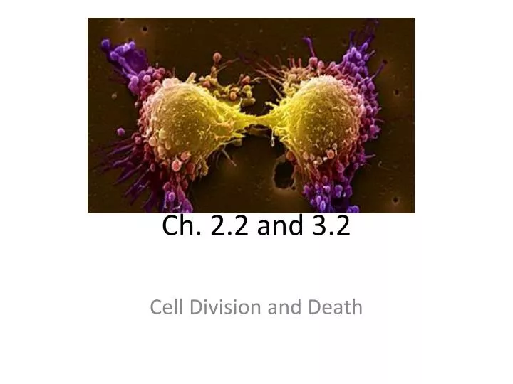
Ch. 2.2 and 3.2. Cell Division and Death. Cell Division & Death. Normal growth and development require an intricate interplay between the rates of two processes Mitosis – Cell division - Produces two somatic cells from one Apoptosis – Cell death

E N D
Ch. 2.2 and 3.2 Cell Division and Death
Cell Division & Death • Normal growth and development require an intricate interplay between the rates of two processes • Mitosis – Cell division - Produces two somatic cells from one • Apoptosis – Cell death - Precise genetically-programmed sequence. EX: Webbing between fingers and toes. Frog Tails.
Cell Division • SOMATIC CELLS (body cells) are made by the process of mitosis. • SEX CELLS (gametes) are made by the process of meiosis.
The Cell Cycle • Broken into two basic time frames: INTERPHASE (not dividing) and MITOSIS (dividing). Figure 2.14
Overview: Stages of the Cell Cycle • Interphase - Prepares for cell division - Replicates DNA and subcellularstructures - Composed of G1, S, and G2 - Cells may exit the cell cycle at G1 or enter G0, a quiescent phase • Mitosis – Division of the nucleus • Cytokinesis– Division of the cytoplasm
INTERPHASE • GAP PHASE I: • Cells normal functioning occurs during G1. The cell resumes synthesis (production) of proteins, lipids, and carbohydrates. • This phase varies drastically among different cell types. EX: Slowly dividing cells (liver cells) may remain in G1 for years. Quickly dividing cells like the bone marrow spend as little as 16 to 24 hours in G1.
SYNTHESIS PHASE: • -This phase includes the REPLICATION of the entire GENOME (DNA) and assembly of proteins, lipids, etc. needed for replication. • This phase takes 8-10 hours in most human cells. • Proteins that will from the spindle structure are made.
Replication of Chromosomes • Process of duplicating a chromosome • Occurs prior to division, during S of interphase • Produces sister chromatids • Held together at centromere Figure 2.14
GAP PHASE II: • -The final preparation for the CELL DIVISION. • - Membranes that were formed in G1 are assembled and stored as empty vesicles beneath the cell membrane. These will be used to enclose both daughter cells. • Some specialized cells go from G1 to G0. They never go through the synthesis or G2 phases. These cells cannot divide, but they can maintain cellular functions.
MITOSIS, the process of CELL DIVISION that produces 2 identical DAUGHTER CELLS from one, occurs in most cells of the body, or SOMATIC CELLS. • -Each DAUGHTER CELL receives the full set of 23 CHROMOSOME PAIRS, just like the PARENT. • MITOSIS begins with the replicated DNA condensed into CHROMOSOME form, with two identical CHROMATIDS attached at a CENTROMERE.
Prophase • In PROPHASE (1st phase) • Replicated chromosomes condense • Microtubules organize into a spindle (centrioles) • Nuclear membrane breaks down
Metaphase • In METAPHASE the SPINDLE FIBERS attach to the CHROMOSOMES at the CENTROMERES and pull them to the cell equator (middle = metaphase plate).
Anaphase In ANAPHASE • Centromeres divide • Chromosomes (chromatids) move to opposite ends of the cell
Telophase & Cytokinesis • In TELOPHASE, the SPINDLES fall apart, the NUCLEOLUS and NUCLEAR MEMBRANES re-form, and • CYTOKINESIS distributes macromolecules and ORGANELLES between the two DAUGHTER CELLS.
If mitosis happens to little, then an injury will go unrepaired, if it happens to often, then an abnormal growth will form. • There are many CHECKPOINTS that control the CELL CYCLE. • Mammalian cells will only divide about 40 – 60 times. A sort of internal “clock” controls the number of divisions.
Cell Cycle Control Proteins called “checkpoint proteins” monitor progression through the cell cycle. Figure 2.16
TELOMERES • Located at the ends of the chromosomes • Contain hundreds to thousands of six nucleotide repeats • Most cells lose 50-200 repeats after each cell division • After about 50 divisions, shortened telomeres signal the cell to stop dividing • Sperm, eggs, bone marrow, and cancer cells produce an enzyme that prevents shortening of telomere • CROWDING limits mitosis.
HORMONES and GROWTH FACTORS can also influence MITOTIC RATES. • CELL DEATH (APOPTOSIS) is as important to body growth and DEVELOPMENT as is MITOSIS. • -When “DEATH RECEPTORS” receive signals to die, enzymes are activated to snip apart cell components in a stepwise cycle. • -PHAGOCYTES gobble up the remains when called in. • -If APOPTOSIS is too infrequent, a CANCER may grow out-of-control.
Sexual Reproduction • Why sexual reproduction? • shuffles alleles; new combinations • provides genetic variation in species
Gametes • Form from cell division of germline cells • Meiosis is cell division to produce gametes • Meiosis has two divisions of the nucleus (Meiosis I and Meiosis II) and produces cells with half the number of chromosomes (haploid) • Whereas SOMATIC CELLS are DIPLOID (containing a double-set of genetic information), the GAMETES are HAPLOID (containing only one copy)
from mother from father child too much! Meiosis • Reduces the genetic material by half • Why is this necessary? meiosis reduces genetic content
Homologous Chromosomes • Matched Pair of Chromosomes = Carry the same genes • Pair during Meiosis I • Separate in the formation of gametes • One copy of each pair is from the mother and one is from the father. Figure 1.2
eye color locus eye color locus hair color locus hair color locus Paternal(fromDad) Maternal(from Mom) Homologous Chromosomes
Sexual Reproduction • Meiosis and sexual reproduction increases genetic diversity in a population • Variation is important in a changing environment • Evolution is the genetic change in a population over time
Meiosis I (reduction division) Meiosis II (equational division) Diploid Haploid Haploid Meiosis: Cell Division in Two Parts Result: one copy of each chromosome in a gamete.
Meiosis Interphase precedes meiosis I Meiosis I Prophase I Metaphase I Anaphase I Telophase I Meiosis II Prophase II Metaphase II Anaphase II Telophase II
Spindle fibers Nucleus Nuclear envelope Prophase I (early) (diploid) Prophase I (late) (diploid) Metaphase I (diploid) Anaphase I (diploid) Telophase I (diploid) Meiosis I : the reduction division
In PROPHASE I, HOMOLOGOUS CHROMOSOME PAIRS line up gene-by-gene in SYNAPSIS (Pairing of homologous chromosomes) and sometimes CROSSING-OVER occurs. • -Each HOMOLOGOUS CHROMOSOME PAIR contains four CHROMATIDS
A A a a B B b b C C c c D D d d E E e e F F f f Recombination (crossing over) • Occurs in prophase of meiosis I • Homologous chromosomes exchange genes • Generates diversity Figure 3.5
Recombination (crossing over) A a a • Exchange between homologs • Occurs in prophase I A B b b B c C C c D D d d E E e e F F f f Figure 3.5 Letters denote genes and case denotes alleles
Recombination (crossing over) a A a A B b B b c c C C • Creates chromosomes with new combinations of alleles for genes A to F D D d d E E e e F F f f Figure 3.5
sister chromatids sister chromatids Tetrad Prophase I - Synapsis Nonsister chromatids
Tetrad nonsister chromatids Chiasma: site of crossing over Crossing Over - Provides Variation variation
In METAPHASE I, the HOMOLOGUES line up in one of 8,388,608 ways and undergo INDEPENDENT ASSORTMENT as HOMOLOGUES are separated in ANAPHASE I.
Independent Assortment The homolog of one chromosome can be inherited with either homolog of a second chromosome. Figure 3.6
Prophase II (haploid) Metaphase II (haploid) Anaphase II (haploid) Telophase II (haploid) Four nonidentical haploid daughter cells Meiosis II : the Equational division Figure 3.4
In METAPHASE II, the CHROMOSOMES line up once again, and in ANAPHASE II, the SISTER CHROMATIDS separate.
TELOPHASE II brings the new NUCLEAR ENVELOPES in, resulting in four HAPLOID CELLS, each carrying a new combination of the GENOME.
Results of Meiosis • Gametes • Four haploid cells • Contain one copy of each chromosome and one allele of each gene • Each cell is unique Figure 3.4