Download
1 / 92
930 likes | 1.21k Views
NUCLEAR MEDICINE & POSITRON EMISSION TOMOGRAPHY. Richard S.Kuebler,M.D.,J.D. Clinical Assistant Professor Academic Director of Nuclear Medicine LSUHSC Department of Radiology Acknowledgements for Lecture and Images Dan Fertel,M.D. Stephanie Casey,M.D.,M.P.H. References. Brant and Helms

E N D
NUCLEAR MEDICINE &POSITRON EMISSION TOMOGRAPHY Richard S.Kuebler,M.D.,J.D. Clinical Assistant Professor Academic Director of Nuclear Medicine LSUHSC Department of Radiology Acknowledgements for Lecture and Images Dan Fertel,M.D. Stephanie Casey,M.D.,M.P.H.
References • Brant and Helms Fundamentals of Diagnostic Radiology • Mettler and Guiberteau Essentials of Clinical Nuclear Medicine
Nuclear Medicine • Therapeutic • Diagnostic
Nuclear Medicine • Use of radioactive isotopes • Most common Technetium-99m • which has a 6 hour half life with good detector • Others include Iodine-123, Indium-111, Gallium-67. • Gamma camera imaging • SPECT imaging
Radioative decay Gamma camera Light pulse Voltage Signal Image Radionuclides= Isotopes attempting to reach stability by emitting radiation Tc-99m ATOM Gamma ray/photon emission (140KeV)
A gamma camera is a device used to image gamma radiation emitting radioisotopes, a technique known as scintigraphy
Uses of Nuclear Medicine • Heart: myocardial perfusion imaging where there has been further development for improvement, rather than decrease or replacement by other modalities. Use SPECT evaluation • Bone scans, evaluate for metastatic bone cancer, osteomyelitis. Most common changes we see are arthritic, correlate with other studies.
Radionuclides for Imaging • Normal Whole-body distribution • Route of excretion • Target/critical organ
Cardiovascular Nuclear Imaging 1. Heart wall motion 2. Myocardial Perfusion & Viability
Heart wall motion(Regional & Global Ventricular Function) • Tc-99m tagged red blood cells • Evaluate left ventricular ejection fraction (nl > 50%) • Tagged red blood cells also used for GI bleeding and hemangioma evaluation in the liver
Equilibrium Radionuclide Angiography Gated equilibrium radionuclide angiograms (MUGA scans) • Performed with Tc-99m red blood cells • Common indications include assessment of LVEF & regional wall motion
Myocardial Perfusion & Viability • Originally thallium 201 • Now use Tc99m Cardiolite or Myoview • SPECT Imaging • Determine adequacy of blood flow to myocardium, especially in conjunction with exercise or pharmacoliogic stress
Myocardial Perfusion • Stress/Exercise • Increased Oxygen demand • Dilate coronary arteries
s ant Apex to base LAD sep Apex lat Cx RCA inf Inf to Ant H V Sep to Lat Normal heart Myocardial Perfusion Rest = baseline perfusion Stress = maximal perfusion
Apical septal Ischemia
Ischemia Inferior Wall
Delayed imaging or PET Viability Fixed defect Scar vs Hibernating myocardium
Fixed defect vs diaphragm Liver, spleen, bowl activity – reconstruction artifact
Breast attenuation Anterior or lateral wall defect
Imaging for infection • Ga 67 Citrate: not used as much anymore, former use was for neoplasm (including lymphoma and lung cancer), inflammation, infection • Ga-67-----complexes with plasma transferrin----carried to sites of inflammation • Incorporated into WBCs—bound by intracellular lactoferrin—then migrate to inflammed areas • Taken up by microorganisms by binding to siderophores produced by bacteria • In-111 tagged white blood cells. Can also tag with Tc-99, but shorter half life.
Gallium scan – Ga-67 • Photon poor- grainy images • Image 24-48 hours • Bowl activity • Sarcoidosis • FUO • Diskitis/spinal Osteomyelitis • Opportunistic infections • Need CXR correlation • PCP – intense activity • Kaposi’s sarcoma – no activity • Normal CXR-Normal Ga-67
In-111 WBC scan • Image 12-24 hr • No bowl/renal activity • Bacterial infections • Prosthetic joint infection – map with Tc-sulfur colloid • Diabetic foot infection
Bone scan Tc99m-MDP • Increased osteoid formation/mineralization of osteoid • Increased blood flow • Can be affected by administed drugs • Always obtain radiographic correlation
Bone Scan - Metastasis • Quality of life • Therapeutic decision making • Multifocal areas increased activity • Red marrow: thorax, ribs, pelvis, limbs, skull
Bone Scan - Metastasis • For a lytic lesion to be visualized by radiography localized demineralization of 30-50% must occur • Bone scans usually demonstrate metastatic lesions much earlier than radiography • False negative bone scan: • Multiple Myeloma • Renal cell carcinoma • Thyroid carcinoma
Bone Scan - Metastasis • 80% of patients with known neoplasms & bone pain have metastasis documented by the bone scan
Flow Blood pool Bone scan 3-phase
Delay Bone scan 3-phase • 3–phase positive: • Osteomyelitis • Acute fractures • Bone tumors • Cellulitis – 2-phase • Shin splints – delay only
Uses of Nuclear Medicine • Thyroid, Iodine-123 or Technitium-99 • Liver-spleen, largely replaced by CT or MRI • Biliary, filling gallbladder, biliary ducts • Renal scan • Brain scans, replaced by CT • Others
Thyroid scan • Thyroid Scan using Tc99 pertechnitate or I-123 • Radioactive Iodine uptake using I-123 or I-131 • Normal uptake in our area 5-15% for 6 hours and 5-25% for 24 hours (10-40% for 24 hours in North and Midwest) • Total body I-131 used for thyroid cancer evaluation
Hypo or hyperfunction Nodules Ectopic thyroid Organification defect Thyroid scan
Total body I-131 • Post-thyroidectomy • Postradioiodine Therapy imaging • I-131: treatment of Graves’ disease and multinodular goiter
Hepatobiliary Imaging (morphine injection) • 1 hr – Gallbladder not visualized
Renal scan Tc-99m MAG3 • Renal function, images similar to IVP • Indications: Renal srtery stenosis • Acute tubular necrosis, obstruction, pyelonephritis
Uses of Nuclear MedicineLymphatic mapping • 99m-Tc Sulfur Colloid • Breast carcinoma and melanoma • Injection for lymph node localization for biopsy (sentinal node)
Liver scan: Tc99 Sulfur colloid • Hepatocellular disease • Confirmation of specific space occupying lesions – ie, focal nodular hyperplasia
Radiology Evaluation of Cancer • Plain Films and Associated Studies • CT Scan • MRI • Nuclear Medicine • Ultrasound • PET
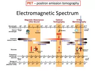
Decay • Positrons • Positron + electron collision • Annihilation reaction generates two 511-keV gamma photons Positron Emission Tomography - PET • Radionuclide with excess protons • PET detector ring for localization & imaging
Type of Pet Scanners • PET Scanner • PET/CT Fusion Scanner
PET – CT Fusion Scanner • Combination of Positron Emission Tomography (PET) and Helical CT • PET detects area of increased metabolic activity as indicated by uptake of radioactive glucose (tumor, infection) • PET data is then “fused” with CT data to produce an image showing increased glucose uptake superimposed upon the exquisite anatomic detail of helical CT
Uses of PET • Brain • Cardiac • Oncology
Cancers evaluated with PET • Lung • Lymphoma • Melanoma • Colorectal • Breast • Esophagus • Head and Neck
Also • Thyroid carcinoma: Approved for history of only 1 type of thyroid carcinoma (Follicular) with negative I-131 scan and rising tumor markers • GU malignancies (Renal, Prostate, Cervical and Ovarian) • Under review for sarcomas • Outpatient procedure
Uses of PET • Diagnosis of cancer (especially lung) • Staging of cancer • Restaging of cancer