Download
1 / 43
440 likes | 549 Views
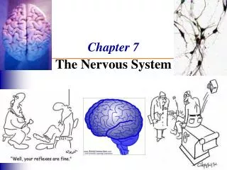
Chapter 7c. The Nervous System. Regions of the Brain: Cerebellum. Two hemispheres with convoluted surfaces Looks like cauliflower Dorsal; under occipital lobe of cerebrum Outer cortex = gray matter; inner cortex = white matter Provides involuntary coordination of body movements

E N D
Regions of the Brain: Cerebellum • Two hemispheres with convoluted surfaces • Looks like cauliflower • Dorsal; under occipital lobe of cerebrum • Outer cortex = gray matter; inner cortex = white matter • Provides involuntary coordination of body movements • Balance & equilibrium • Impulses from ear, eye, touch receptors of skeletal muscles & tendons • Ataxia – damage to cerebellum; loose balance, become clumsy
Cerebellum Figure 7.15a
Protection of the Central Nervous System • Scalp and skin • Skull and vertebral column • Meninges • Cerebrospinal fluid (CSF) • Blood-brain barrier Figure 7.16a
Protection of the Central Nervous System Figure 7.17a
Meninges • 3 connective membranes • Dura mater • Leathery = hard mother • Double-layered external covering • Periosteum – attached to inner surface of the skull • Meningeal layer – outer covering of the brain • Folds inward in several areas to attach to cranial cavity
Meninges • Arachnoid layer = weblike = spider • Middle layer • Web-like – span the subarachnoid space to attach to pia matter • Pia mater = gentle mother • Internal layer • Clings to the surface of the brain
Meninges Figure 7.17b
Cerebrospinal Fluid (CSF) • In subarachnoid space • Similar to blood plasma composition • Formed by the choroid plexus – capillaries hanging from roof of ventricles • Forms a watery cushion to protect the brain • Circulated in arachnoid space, ventricles, and central canal of the spinal cord • Absorbed into venous blood in dural sinuses
Ventricles and Location of the Cerebrospinal Fluid Figure 7.17a–b
Ventricles and Location of the Cerebrospinal Fluid Figure 7.17c
Meningitis – inflammation of meninges • Bacterial or viral • CSF – similar to plasma • Less protein, more vitamin C • Hydrocephalus = water on brain • Obstruction of CSF drainage
Hydrocephalus in a Newborn • Hydrocephalus • CSF accumulates and exerts pressure on the brain if not allowed to drain Figure 7.19
Blood Brain Barrier • Includes the least permeable capillaries of the body • Excludes many potentially harmful substances • Brain could not handle fluctuations of chemicals in blood • Only water, glucose, essential amino acids can pass in • Useless as a barrier against some substances • Fats and fat soluble molecules • Respiratory gases • Alcohol • Nicotine • Anesthesia
Traumatic Brain Injuries • Brain injured at site of blow & effect of ricocheting & hitting opposite end of skull • Concussion (see stars) • Slight brain injury • No permanent brain damage • Contusion • Nervous tissue destruction occurs (cerebral cortex injury – conscious; brain stem injury – coma) • Nervous tissue does not regenerate • Cerebral edema or intracranial hemorrhage • Swelling from the inflammatory response • May compress and kill brain tissue
Cerebrovascular Accident (CVA) • Commonly called a stroke • 3rd leading cause of death • The result of a ruptured blood vessel supplying a region of the brain or a blood clot • Brain tissue supplied with oxygen from that blood source dies • Loss of some functions or death may result • Aphasias- • Motor – damage to Broca’s area; can’t talk • Sensory – can’t understand written or spoken language • Some recovery – undamaged neurons spread into damaged area
Alzheimer’s Disease • Progressive degenerative brain disease • Mostly seen in the elderly, but may begin in middle age • Structural changes in the brain include abnormal protein deposits and twisted fibers within neurons • Decrease in Ach, gyri shrink, brain atrophies • Victims experience memory loss, irritability, confusion and ultimately, hallucinations and death
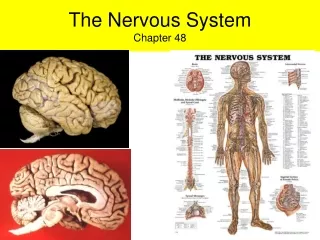
Spinal Cord • Extends from the foramen magnum of the skull (from medulla oblongata) to the region of T12, L1 • 31 pair of spinal nerves arise from the spinal cord • 17” long • 2-way conduction pathway to brain Figure 7.18
Below T12 is the cauda equina (a collection of spinal nerves) • Because vertebrae grow faster than cord, cord doesn’t reach end of vertebral column • Enlargements occur in the cervical and lumbar regions • Serve upper & lower limbs • Major reflex center • Covered by meninges
Spinal Cord Anatomy Figure 7.20 (1 of 2)
Spinal Cord Anatomy Figure 7.20 (2 of 2)
Spinal Cord Anatomy • Internal gray matter - mostly cell bodies • Dorsal (posterior) horns – association or interneurons & sensory neurons • Anterior (ventral) horns – motor neurons of somatic (voluntary) • Gray matter surrounds the central canal • Central canal is filled with cerebrospinal fluid • Exterior white matter – conduction tracts • Dorsal, lateral, ventral columns • Dorsal & ventral roots fuse into spinal nerves • Central Canal filled with cerebrospinal fluid Figure 7.19
Spinal Cord Anatomy Figure 7.21
Spinal Cord Anatomy • Meninges cover the spinal cord • Spinal Nerves leave at the level of each vertebrae • Dorsal root – cell bodies of sensory neurons • Associated with the dorsal root ganglia – collections of cell bodies outside the central nervous system • Ventral root – motor neurons of somatic system • Contains axons
Peripheral Nervous System (PNS) • Nerves and ganglia (=groups of neurons cell bodies) outside the central nervous system • Nerve = bundle of neuron fibers • Neuron fibers are bundled by connective tissue
PNS: Structure of a Nerve • Endoneurium surrounds each fiber • Groups of fibers are bound into fascicles by perineurium • Fascicles are bound together by epineurium Figure 7.20
PNS: Structure of a Nerve Figure 7.23
PNS: Classification of Nerves • Mixed nerves • Both sensory and motor fibers (all spinal nerves are mixed) • Sensory (afferent) nerves • carry impulses toward the CNS • Motor (efferent) nerves • carry impulses away from the CNS
PNS: Cranial Nerves • 12 pairs of nerves that mostly serve the head and neck • Only the pair of vagus nerves extend to thoracic and abdominal cavities • Numbered in order, front to back • Most are mixed nerves, but three are sensory only • Optic, olfactory, vestibulocochlear
PNS: Cranial Nerves • I Olfactory nerve – sensory for smell • II Optic nerve – sensory for vision • III Oculomotor nerve – motor fibers to eye muscles • IV Trochlear – motor fiber to eye muscles
Cranial Nerves • V Trigeminal nerve – sensory for the face; motor fibers to chewing muscles • VI Abducens nerve – motor fibers to eye muscles • VII Facial nerve – sensory for taste; motor fibers to the face • VIII Vestibulocochlear nerve – sensory for balance and hearing
Cranial Nerves • IX Glossopharyngeal nerve – sensory for taste; motor fibers to the pharynx • X Vagus nerves – sensory and motor fibers for pharynx, larynx, and viscera • XI Accessory nerve – motor fibers to neck and upper back • XII Hypoglossal nerve – motor fibers to tongue
PNS: The Cranial Nerves Table 7.1 (1 of 4)
PNS: The Cranial Nerves Table 7.1 (2 of 4)
PNS: The Cranial Nerves Table 7.1 (3 of 4)
PNS: The Cranial Nerves Table 7.1 (4 of 4)
PNS: Distribution of Cranial Nerves Figure 7.24