Download
1 / 20
200 likes | 459 Views
Mechanisms of Bcl-2 in Programmed Cell Death. Laura Beth Hill St. Edward’s University. Apoptosis. Essential for normal embryonic development Natural and pathological Morphologic characteristics Regulated by proteins in Bcl-2 family. The Apoptotic Process. Cell receives death signal

E N D
Mechanisms of Bcl-2 in Programmed Cell Death Laura Beth Hill St. Edward’s University
Apoptosis • Essential for normal embryonic development • Natural and pathological • Morphologic characteristics • Regulated by proteins in Bcl-2 family
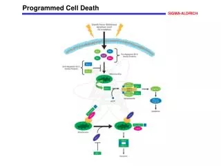
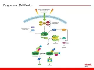
The Apoptotic Process • Cell receives death signal • Mitochondrial membrane potential decreases • Transport of cytochrome c through membrane into cytosol • Cytochrome c binds to Apaf-1 • Caspase activity initiated • Cell degradation
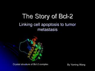
What is Bcl-2? • Family of proteins that includes promoters and inhibitors • Proto-oncogene • Localized to outer mitochondrial membrane • Can form homo- and heterodimers • Exhibits biphasic expression
FOCUS OF SEMINARPossible Mechanisms • Bcl-2 blocks release of cytochrome c from mitochondrial membrane (Yang, et al., 1997) • Bcl-2 forms channels in lipid membranes (Schendel, et al., 1997)
Cytochrome c Model • Necessary for the initiation of apoptosis • Found in the mitochondrial intermembrane space • Localization suggests connection between Bcl-2 and cytochrome c
HL-60cells neo cells bcl-2 cells Isolation Isolation Staurosporine Staurosporine Immunoblot analysis Immunoblot analysis
Yang’s Results • Cytochrome c in neo cells showed cytosol increase, with corresponding decrease in mitochondria • No significant change of cytochrome c in mitochondria or cytosol of Bcl-2 cells • No significant amount of cytochrome c found in cytosol of control cells
Yang’s Conclusion • Bcl-2 prevents the release of cytochrome c • Mechanism by which Bcl-2 blocks release unknown • Structural similarity to bacterial toxins suggests pore-forming ability
Channel Formation Model • Bcl-2 can regulate ionfluxes and protein translocation • 3D structure of Bcl-xL is similar to the pore-forming domains of DT and the bacterial colicins
Protein Production Bcl-2 wild-type Bcl-2 (5,6) mutant Cl- efflux assay Cl- efflux assay Detection of single channels Detection of single channels
Schendel’s Results • Bcl-2 formed ion-conducting pores in a manner similar to that of bacterial toxins • Bcl-2 mutant produced only non-specific Cl- efflux • Bcl-2 in planar lipid bilayers formed discrete cation-selective channels • Bcl-2 mutant did not form channels here
Schendel’s Conclusions • Biophysical evidence proves that Bcl-2 forms channels in membranes • Channels reside in closed state • What regulates channels? • How do pro-apoptotic proteins oppose anti-apoptotic proteins?
Practical Importance • Association of disease with inappropriate apoptosis • Adjustment of apoptotic threshold • Gene therapy to control cell death • Protection of developing nervous system against neurotoxins (e.g. EtOH)
Acknowledgements St. Edward’s School of Natural Sciences faculty and staff