Download
1 / 94
980 likes | 1.27k Views
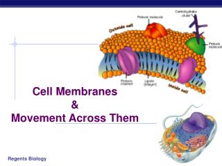
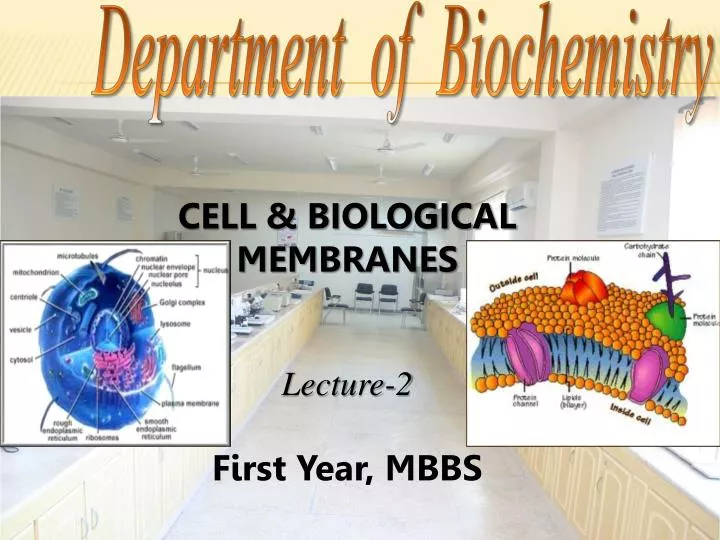
Department of Biochemistry. CELL & BIOLOGICAL MEMBRANES Lecture-2 First Year, MBBS. Learning Objectives By the end of this Lecture, students shall be able to: 1 . Describe the fluid Mosaic Model of cell membrane

E N D
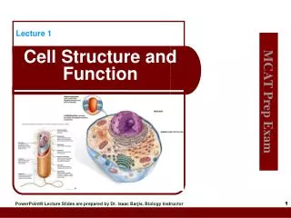
Department of Biochemistry CELL & BIOLOGICAL MEMBRANES Lecture-2 First Year, MBBS
Learning Objectives By the end of this Lecture, students shall be able to: 1. Describe the fluid Mosaic Model of cell membrane 2. Describe the various types of transport across the cell membrane with examples3. Differentiate between the passive and active transport across cell membrane
1. Phospholipids form bilayer. 2. Certain lipids determine fluidity of plasma membrane: Cholesterol Saturated and unsaturated fatty acids (components of phospholipids) 3. Provide permeability barrier for water soluble molecules. FUNCTIONS OF MEMBRANE LIPIDS
4. Provide a hydrophobic region in which part or major part of membrane proteins are embedded. 5. Provide site for attachment of : Peripheral proteins (electrostatic interactions) Oligosaccharide chains FUNCTIONS OF MEMBRANE LIPIDS
Many of them have negative electrical charge which gives most cells an overall negative surface charge that repels other negative objects. The glycocalyx of some cells attaches to the glycocalyx of other cells, thus attaching cells to one another. Carbohydrates play an important role in cell-cell recognition. Many of these act as receptor substance for binding hormones. FUNCTIONS OF MEMBRANE CarboHydrates
Cell-cell recognition: The ability of a cell to distinguish one type of neighboring cell from another. • Cell-cell recognition is crucial in the functioning of an organism. It is the basis for: • Sorting of cells into tissues and organs in an animal embryo’s cell. • Rejection of foreign cells by the immune system. Membrane carbohydrates are important for cell-cell recognition
The way cells recognize other cells is probably by keying on surface molecules (markers) Markers: Surface molecules found on the external surface of the plasma membrane that distinguish one cell from another.
Many of the integral proteins provide structural channels (or pores) through which water molecules & water soluble substances, especially ions, can diffuse b/w the ECF & ICF. These protein channel also have selective properties that allow preferential diffusion of some substances over others e.g. Aquaporins. Other integral proteins act as carrier proteins for transporting substances that otherwise could not penetrate the lipid bilayer. FUNCTIONS OF MEMBRANE PROTEINS: Integral Membrane Proteins
The temperature above which the paracrystalline solid changes to fluid is called Tm. Transition temperature is characteristic for each membrane and depends upon membrane lipid composition. TRANSITION TEMPERATURE (Tm)
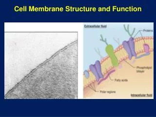
THE FLUID MOSAIC MODEL • Fluid = always moving and changing • Mosaic = made up of many different parts Fluid mosaic model was proposed in 1972 by Singer and Nicolson. According to this model, membrane proteins are like icebergs floating in a sea of phospholipid molecules.
Later on, it was demonstrated that phospholipids also undergo rapid redistribution in the plane of membrane. In FREEZE-FRACTURE TECHNIQUE, cells are frozen to very cold temp and then fractured with a very fine diamond knife. Some cells are fractured between two layers of membrane lipid bilayer. When viewed with Electron microscope, the membrane appeared to be a mosaic, studded with proteins. Due to fluidity of membranes and the appearance of proteins, the concept of membrane structure is called “the fluid mosaic structure”.
Fatty acyl chains in the in the interior of membrane form a fluid, hydrophobic region. Integral proteins float in this sea of lipids held by hydrophobic interactions with their non-polar amino acid side chains. Both lipids and proteins are free to move laterally in the plane of bilayer, but movement of either from one face of the bilayer to the other (flip-flop movement) is restricted. Carbohydrate moieties attached to some proteins and lipids of the plasma membrane are exposed on the extracellular side.
Molecules rarely flip transversely (flip-flop) across the membrane, because hydrophilic parts would have to cross the membrane’s hydrophobic core.
2. TEMPERATURE: Individual hydrocarbon chains of fatty acids are in constant motion. • At low temperature relatively little lipid motion occurs & bilayer exists as a nearly crystalline array. • Above a certain temperature lipid can under go a rapid motion. Fluidity of memb effects its functions: As fluidity increases permeability of membrane to water & other hydrophilic substances increases.
Rat hepatocyte is one of the most extensively studied of all cells from a biochemical viewpoint. This is due to following reasons: Available in relatively large amounts Diverse functions Suitable for fractionation studies; contains major organelles (nucleus, mitochondria, ER, free ribosomes, Golgi-apparatus, lysosomes, peroxisomes, plasma membrane, cytoskeletal elements) found in eukaryotic cells. Study of Cells from A Biochemical Viewpoint
In order to study the function of any organelle, it is necessary to isolate it in relatively pure form. The usual process by which this is achieved is called subcellular fractionation. Subcellular fractionation generally entails three procedures: Extraction Homogenization Centrifugation SUBCELLULAR FRACTIONATION
As a first step toward isolating a specific organelle (or molecule); it is necessary to extract it from the cells in which it is located. Most organelles & many biomolecules are labile & subject to loss of biologic activities; they must be extracted using mild conditions (i.e. employment of aqueous solution, avoidance of extreme of pH, osmotic pressure, high temperature). Most procedures for isolating organelles are performed at 0-4 •C (e.g. In a cold room or using material kept on ice). EXTRACTION
Significant losses of activity occur at room temperature, partly owing to the action of various digestive enzymes (protease, nuclease etc) liberated when cells are disrupted. A common solution for extraction of organelles consist of sucrose, 0.25 mol/l (isoosmotic), adjusted to pH 7.4 by TRIS HCL buffer, 0.05 mol/l, containing K+ & Mg++ ions at near physiologic concentrations; this solution is conveniently called STKM. Organic solvents are used for extraction of lipids & nucleic acids.
Organs (e.g. liver) & their contained cells may be conveniently disrupted by the process of homogenization, in which a manually operated or a motor driven pestle is rotated within a glass tube of suitable dimensions containing minced fragments of the organs under study, & a suitable homogenizing medium, such as STKM. The controlled rotation of the pestle exerts mechanical shearing forces on cells & disrupts them, liberating their constituents in sucrose. The resulting suspension, containing many organelles, is known as HOMOGENATE. HOMOGENIZATION
Subfractionationof the contents of homogenate is done by differential centrifugation. In the classic method, a series of 03 different centrifugation steps at successively greater speed yield a pellet & supernatant. The supernatant from each step is subjected to centrifugation in the next step. This procedure provides three pellets, named the nuclear fraction, mitochondrial fraction, & microsomal fraction. CENTRIFUGATION
NOTE: None of these fractions are composed of absolutely pure organelles. However, it has been established by use of electron microscope & suitable markers (enzymes or chemical components) that the major constituents of each of these 03 fractions are nuclei, mitochondria & microsomes respectively. Nuclear fraction → contain nuclei & unruptured cells Mitochondrial fraction → contain mitochondria, lysosomes & peroxisomes Microsomal fraction → contain mixture of free ribosomes, smooth ER rough ER
A marker enzyme or chemical is one that is almost exclusively confined to one particular organelle. The marker thus can serve to indicate the presence or absence of the organelle in any particular fraction in which it is contained. MARKER ENZYME OR CHEMICAL
TRANSPORTATION THROUGH MEMBRANE: Can occur by following mechanisms: 1. Cross membrane movement of small molecules. • Diffusion • Active transport 2. Cross membrane movement of large molecules. • Endocytosis • Exocytosis 3. Signal transmission across membrane. • Cell surface receptors 4. Inter cellular contact and communication.
TRANSPORT SYSYTEM Transport system can be described in a functional sense according to the no. of molecules moved & direction of movement or according to whether movement is towards or away from equilibrium. Uniport system: Moves one type of molecules bidirectionally. Co-Transport system: The transfer of one solute depends upon the stoichiometric simultaneous transfer of another solute.
Symport: Move these solute in the same direction Examples: Na+ - sugar transporters (glucose & certain other sugars) and Na+- amino acid transporters in mammalian cells • Antiport: Movement of two molecules in opposite direction Examples: Na+ in & Ca2+ out
Uniport Symport Antiport Cotransport
1. Cross membrane movement of small molecules DIFFUSION: • Simple • Facilitated Molecules can passively traverse the bilayer down electrochemical gradients by simple diffusion or by facilitated diffusion.
Concentration gradient across the membrane. The electrical potential across the membrane. The permeability coefficient of the substance for the membrane. The hydrostatic pressure gradient across the membrane. Temperature. Factors Affecting Net Diffusion Of A Substance Through Membrane
The permeability coefficient of the substance is the solubility of that substance in the hydrophobic core of the membrane bilayer.
SIMPLE DIFFUSION Definition:Movement of molecules/solutes from the region of higher solute concentration to the region of lower solute concentration through the membrane. Examples: Non polar gases such as CO2, O2 , nitrogen, methane & alcohol. It can occur by two path ways 1. Through the interstices of the lipid bilayer, if the diffusing substances is lipid soluble. 2. Through watery channels that penetrate all the way through some of large transport proteins.
Solubility is inversely proportionate to the number of hydrogen bonds that must be broken in order for a solute in external aqueous phase to become incorporated in the hydrophobic bilayer. Electrolytes, poorly soluble in lipids, do not form H- bonds with water, but they do acquire a shell of water from hydration by electrostatic interactions.
Size of shell is directly proportional to the charge density of the electrolyte. Electrolytes with a Large charge density have a larger shell of hydration and thus a slower diffusion rate. Example: Na+ has a higher charge density than K+ so K+ move easily through the membrane.
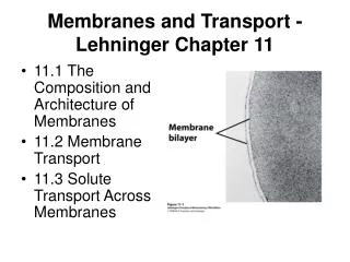
Channels and pores facilitate translocation of molecules or ions across cell membrane by creating a central aqueous channel in the protein that permits diffusion of substrate in both directions. Channel proteins do not bind or sequester the molecule or ions in transit. CHANNELS AND PORES
Their specificity is based on the size and charge of the substance. Cation conductive channels are negatively charged within the channel. The specific channels for Na+, k+ Ca2+ and Cl- have been identified.
Helical region Channel Lipid bilayer _ _ K+
These are regulated by various mechanisms that open or close the passageway. Channels are open transiently and thus are gated. Ligand-gated channels: Binding of ligand to receptor opens the channel. Voltage-gated channel: Open in response to a change in membrane potential.
Despite polarity, water crosses some membranes slowly by simple diffusion due to high concentration gradient. However, for tissues in which rapid transmembrane water movement is essential e.g. Kidneys, water diffuses through channels formed by specific integral proteins – the aquaporins. DIFFUSION OF WATER
Aquaporins are small hydrophobic integral membrane proteins that contain water pore. The aqueous pathway is lined with few hydrophilic residues that attract water. In addition to water, aquaporins permit translocation of CO2 , glycerol, urea, purines, pyrimidines and nucleosides. Eleven (11) mammalian AQPs have been identified. AQUA PORINS (AQP)
These are subdivided by amino acid sequence and functional characteristics into Aquaporins: channel selective only for water Aqua glyceroporins: permit translocation of water and small solutes Aquaporins are present in different tissues. Kidneys (proximal tubules and collecting ducts) contain AQP1, AQP2, AQP3, AQP4, and AQP6.
AQP2 is under hormonal control. Low levels of AQP2 and polyuria are found in acquired nephrogenic diabetes insipidus, acquired hypokalemia and hypercalcemia. High levels of AQP2 are found in congestive heart failure, liver cirrhosis and pregnancy, leading to an expansion of the ECF volume CLINICAL CORRELATION
Transmembrane passage of polar compounds & ions is made possible by membrane proteins that lower the activation energy for transport by providing the alternative path for specific solute through the lipid bilayer. These protein are not enzymes but are called transporters. Specific solutes diffuse down electrochemical gradient across membrane more rapidly than might be expected from their size, change or partition coefficients through facilitated diffusion. FACILITATED DIFFUSION:
In this model the carrier protein exist in two principal conformations. In the “pong” state it is exposed to high concentration of solute. Solute molecules bind to specific sites on the carrier protein. Conformational change occur that exposes the carrier protein to a lower concentration of solute “ping” state and helps transporting solute molecules. PING PONG MECHANISM
pong ping