Download
1 / 33

E N D
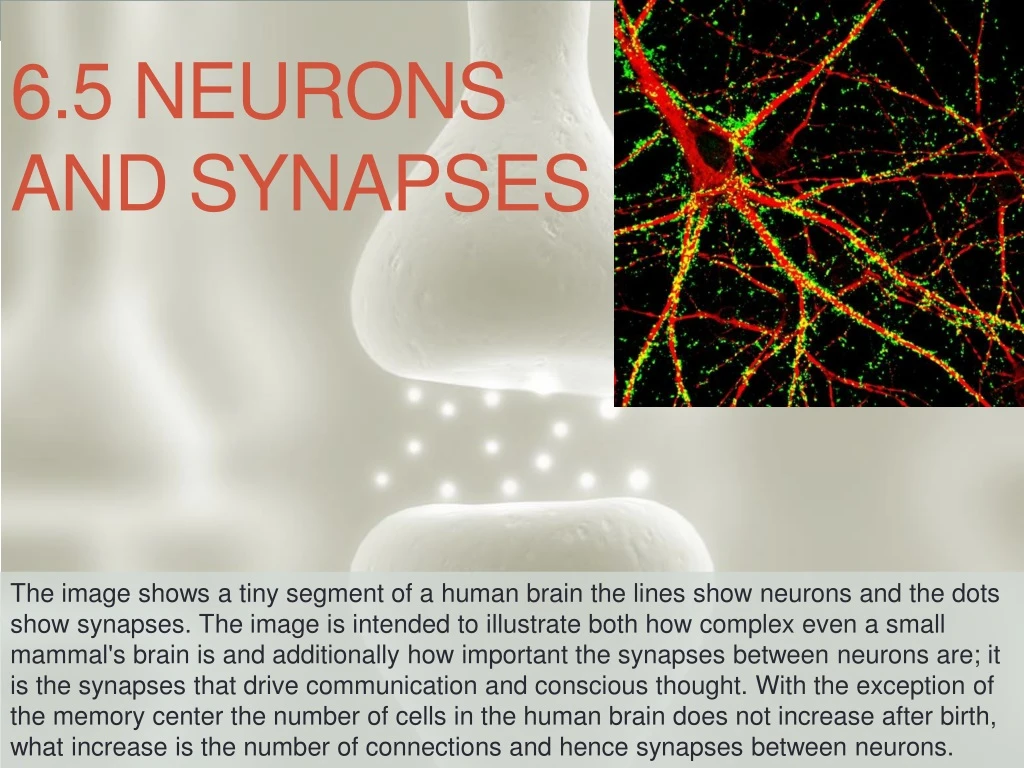
6.5 Neurons and synapses The image shows a tiny segment of a human brain the lines show neurons and the dots show synapses. The image is intended to illustrate both how complex even a small mammal's brain is and additionally how important the synapses between neurons are; it is the synapses that drive communication and conscious thought. With the exception of the memory center the number of cells in the human brain does not increase after birth, what increase is the number of connections and hence synapses between neurons.
6.5 • Essential idea: Neurons transmit the message, synapses modulate the message.
What would it be like to feel no pain? • https://youtu.be/m5B20VvzWqA
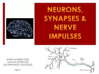
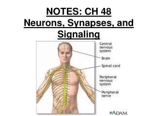
6.5.U1 Neurons transmit electrical impulses. • The Nervous System allows organisms to respond to external and internal stimuli • It consists of: • – Central Nervous System (CNS) • – Peripheral Nervous System (PNS) • Neurons – functional unit of the nervous system; specialized cells for transmitting electrical and chemical signals
Anatomy of a Nerve Cell: • Cell body – contains nucleus, most of the cytoplasm and most of the organelles • Dendrites and axon extend from the cell body • Dendrites – short and highly branched Dendrites Cell body
Axon – conducts impulses away from the cell body to another neuron or to a muscle or gland • Microscopic in diameter but may extend a meter or more in length • – in • Synaptic terminals release (chemicals) (gap between neurons)
Myelin Sheath – fatty material surrounding the axons of neurons of the PNS; • Nodes of Ranvier –
In the PNS: • Nerves consist of • are usually grouped together in masses called • In the CNS: • Bundles of are called or instead of nerves • Collection of are called
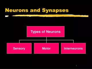
Types of Neurons: • Sensory ( ) neurons – conduct impulses into CNS from the periphery (sensory impulses) • Pick up stimulus from sensory receptors – mechanoreceptors, chemoreceptors, thermoreceptors, photoreceptors
Interneurons ( ) – afferent neurons usually transmit impulses to interneurons • Located within CNS • Neurons that integrate input and output • involves sorting and interpreting incoming sensory information and determining the appropriate response • Forms connecting lines between sensory and motor neurons Brain & Spinal cord
Motor ( ) neurons – transmit messages from CNS to effectors (muscle or gland) Sensory receptors, afferent and efferent neurons are part of the Peripheral Nervous System Afferent (input – “inform” CNS of changing conditions) Efferent (output – transmit the “decisions” of the CNS) http://www.siumed.edu/~dking2/ssb/neuron.htm#4b
PNS Motor Division Autonomic NS :
6.5.U3 Neurons pump sodium and potassium ions across their membranes to generate a resting potential. Membrane potential is the difference in electrical charge across the plasma membrane • Resting potential is the potential difference across a nerve cell membrane when it is not being stimulated. • Slight the membrane and slight the membrane • More negatively charged inside the cell compared to the interstitial fluid outside • Resting potential is normally about -70 millivolts (mV) • Membrane of neuron is – as a result, the cell can produce an action potential(nerve impulse) Resting Potential
6.5.U3 Neurons pump sodium and potassium ions across their membranes to generate a resting potential. • Na+ concentration is 10x greater outside the cell and K+ concentration is 10x greater inside the cell • K+ tends to leak out by diffusion through ion channels causing further negative charge inside as compared to outside of cell • Ion channels that allow the passage of Na+ are closed at resting potential Closed at rest
6.5.U4 An action potential consists of depolarization and repolarization of the neuron. Action Potential • Depolarization is a • Action potential (nerve impulse) is the , reversing the resting potential from about -70 mV to +40 mV. • Repolarization is when the .
6.5.U5 Nerve impulses are action potentials propagated along the axons of neurons. AND 6.5.U9 A nerve impulse is only initiated if the threshold potential is reached. Action Potential • Stimulation – all or none response • Threshold stimulation – • Causes Na+ ion channels to open allowing Na+ to rush into interior of cell (depolarization) • Disturbs adjacent areas – Na+ channels open causing a depolarization wave (nerve impulse) • Polarity across membrane is momentarily reversed • K+ channels also open but more slowly allowing repolarization
6.5.U4 An action potential consists of depolarization and repolarization of the neuron. • Repolarization – after action potential passes, membrane begins to repolarize • Na+ channels close and membrane becomes • Open K+ channels allow • Impulse is actually a series of depolarization and repolarization waves sweeping down the axon (takes place in less than 1 millisecond) • Then K+ channels close and • For every three sodium ions pumped out, two potassium ions are pumped in Impulse conduction video
6.5.U6 Propagation of nerve impulses is the result of local currents that cause each successive part of the axon to reach the threshold potential. Propagation of a nerve impulse in un-myelinated axons Cell body http://cnx.org/resources/0d4d8e978090c5adf07cc1661372b69be3496ec6/Figure_35_02_04.png
6.5.U6 Propagation of nerve impulses is the result of local currents that cause each successive part of the axon to reach the threshold potential. Propagation of a nerve impulse in un-myelinated axons http://highered.mheducation.com/olcweb/cgi/pluginpop.cgi?it=swf::535::535::/sites/dl/free/0072437316/120107/bio_d.swf::Action%20Potential%20Propagation%20in%20an%20Unmyelinated%20Axon
6.5.S1 Analysis of oscilloscope traces showing resting potentials and action potentials. Na+/K+ pump Na+& K+channels closed Na+ channels open Inside of cell becomes positive Na+ channels close K+channels open Negative charge restored inside Na+/K+ pump Na+ & K+ channels closed
6.5.U2 The myelination of nerve fibers allows for saltatory conduction. • Myelinated vs. Non-myelinated • Impulse conduction is • Here there is continuous conduction, the entire axon must depolarize • – speeds up transmission • Depolarization occurs only at the Nodes of Ranvier – Action Potential “jumps” from one node to the next (“to leap”) • Degradation of the impulse is reduced and allows the impulse to travel longer distances • as the quantity of sodium and potassium ions that need to be pumped to restore resting potential is less than that of a un-myelintated axon • Diameter of axon also affects speed of transmission • Larger diameters transmit faster *The jump along the axon is actually just the very rapid conduction inside the myelinated portion of the axon. Myelinated vs. non-myelinated animation: http://www.wiley.com/college/jenkins/0470227583/animations/ch12/nerve3a/screen_3_2.swf
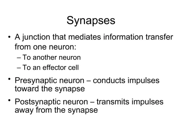
6.5.U7 Synapses are junctions between neurons and between neurons and receptor or effector cells. • Synapse (synaptic cleft or gap) – • Synapse between neuron and muscle cell is called a neuromuscular junction or motor end plate
6.5.U8 When presynaptic neurons are depolarized they release a neurotransmitter into the synapse. AND 6.5.U9 A nerve impulse is only initiated if the threshold potential is reached.
6.5.U8 When presynaptic neurons are depolarized they release a neurotransmitter into the synapse. AND 6.5.U9 A nerve impulse is only initiated if the threshold potential is reached.
6.5.A1 Secretion and reabsorption of acetylcholine by neurons at synapses. Acetylcholine is a neurotransmitter used in many synapses through the nervous system One use is at the neuromuscular junction, i.e. it is the molecule that motor neurons release to activate muscles. Interfering with the action of acetylcholine can cause a range of effect from paralysis to convulsions. http://faculty.pasadena.edu/dkwon/chap%208_files/images/image61.png
6.5.A2 Blocking of synaptic transmission at cholinergic synapses in insects by binding of neonicotinoid pesticides to acetylcholine receptors.
6.5.A2 Blocking of synaptic transmission at cholinergic synapses in insects by binding of neonicotinoid pesticides to acetylcholine receptors.
6.5.A2 Blocking of synaptic transmission at cholinergic synapses in insects by binding of neonicotinoid pesticides to acetylcholine receptors.
Nature of science: Cooperation and collaboration between groups of scientists - biologists are contributing to research into memory and learning. (4.3) Nowadays scientists often work in multidisciplinary teams for example the Centre for Neural Circuits and Behaviour (CNCB) The aim of the CNCB is to understand how intelligence emerges from the physical interaction of nerve cells. Studying the brain from this top-down approach to answer such fundamental questions requires techniques and understanding from a range of disciplines. GeroMiesenböck FRS Waynflete Professor of Physiology, Wellcome Investigator Martin Booth Professor of Engineering Science Tim Vogels Sir Henry Dale Fellow (physicsist) Scott Waddell Professor of Neurobiology, Wellcome Trust Senior Research Fellow in Basic Biomedical Sciences Stephen Goodwin Professor of Neurogenetics, Wellcome Investigator Korneel Hens Group Leader (Biochemist) http://www.cncb.ox.ac.uk/team/