Download
1 / 75
870 likes | 1.32k Views
Protein folding in the cell. Basics - cell compartments, molecular crowding: cytosol, ER, etc. Folding on the ribosome - co-translational protein folding Molecular chaperones - concepts , introduction - intramolecular chaperones - chemical chaperones - protein chaperones.

E N D
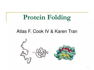
Protein folding in the cell Basics - cell compartments, molecular crowding: cytosol, ER, etc. Folding on the ribosome - co-translational protein folding Molecular chaperones - concepts, introduction - intramolecular chaperones - chemical chaperones - protein chaperones
Cell compartments and folding • eukaryotes - cytosol ..................................protein synthesis, folding/assembly - extracellular .........................proteins are exported in folded form - mitochondria ........................limited protein synthesis; energy production - chloroplasts ..........................limited protein synthesis; light harvesting - endoplasmic reticulum.......... import of unfolded proteins; protein processing - peroxisome ........................... import of folded proteins; anab./catab. pathways - nucleus ................................. import of folded proteins - lysosome................................ import of unfolded proteins; degradation • bacteria - cytosol ..................................protein synthesis, etc. - periplasm .............................import and folding of periplasmic proteins - extracellular .........................proteins are exported • archaea - cytosol ..................................protein synthesis, etc. - extracellular .........................proteins are exported
Folding in vitro vs.in vivo in vitro in vivo protein denatured in a chaotrope Differences: 1. One has all of the information immediately available for folding; the other process is gradual 2. the cellular environment is very different (much more crowded) folding by dilution in buffer folding folded protein folded protein
Co-translational protein folding Fact: - first ~30 amino acids of the polypeptide chain present within the ribosome is constrained (the N-terminus emerges first) Assumption: as soon as the nascent chain is extruded, it will start to fold co-translationally (i.e., acquire secondary structures, super-secondary structures, domains) until the complete polypeptide is produced and extruded folding assembly
Sindbis Virus Capsid Protein (SCP) catalytic triad & C-terminus of SCP • SCP is the capsid protein of the Sindbis virus • 26S Sindbis RNA encodes a polyprotein • SCP is auto-proteotically cleaved from the rest of the polyprotein • other cellular proteases cleave E1-E3 from the polyprotein to generate the mature proteins; E1, the envelope protein, is 9 kDa • SCP is a 33 kDa serine protease • WT SCP self-cleaves; Ser215 => Ala215 mutant doesn’t N C SCP E1 E2 E3
SCP folds co-translationally 2 3 4 5 6 7 8 10 12 min Result: Mut SCP-E1 42 kDa 33 kDa 9 kDa 2 3 4 5 6 7 8 10 12 min 42 kDa WT SCP 33 kDa 9 kDa 2 3 4 5 6 7 8 10 12 min WT SCP- E1 42 kDa 33 kDa 9 kDa Experiment: 1. make and translate different SCP construct RNAs in vitro in the presence of 35S-methionine for 2 min 2. Prevent re-initiation of translation with aurintricarboxylic acid (ATCA): ‘synchronizing’ 3. at set timepoints, add SDS buffer and perform SDS-PAGE 4. observe by autoradiography * N C SCP E1 N C SCP N C SCP E1
Macromolecular crowding E. coli cytosol ~340 mg/ml ribosome proteins other macromolecules chaperonin nucleic acids in vitro <0.1 mg/ml Ellis and Hartl (1996) FASEB J. 10:20-26 When doing experiments in vitro, we should all be thinking about this:proteins in isolated (pure) systems may not behave as they do in the cell- binding partner(s) might be missing - cell conditions (pH, salts, etc.- post-translational modifications might be missing may be dramatically different
Effects of crowding Definition: Molecular crowding is a generic term for the condition where a significant volume of a solution, or cytoplasm for example, is occupied with things other than water Fact: - association constants (ka) increase significantly - dissociation constants (kd) decrease significantly (kd=1/ka) - increased on-rates for protein-protein interactions (see for example Rohwer et al. (2000) J. Biol. Chem.275, 34909) Assumption: - non-native polypeptides will have greater tendency to associate intermolecularly, enhancing the propensity of aggregation
Effects of crowding: example oxidized lysozyme reduced lysozyme denatured lysozyme, reduced or oxidized dilution in buffer with different crowding agents loss of activity due to protein aggregation measure lysozyme activity crowding agents: ficoll 70*, dextran 70, protein (BSA, ovalbumin) *roughly spherical polysaccharide van den Berg et al. (1999) EMBO J. 18, 6927.
Problem: non-native proteins exposed hydrophobic residues X X X X incorrect molecular interactions & loss of activity intramolecular intermolecular X X X X X X misfolding aggregation • non-native proteins expose hydrophobic residues that are normally buried within the ‘core’ of the protein • these hydrophobic amino acids have a strong tendency to interact with other hydrophobic (apolar) residues - especially under crowding conditions
Solution: molecular chaperones • in the late 1970’s, the term molecular chaperone was coined to describe the properties of nucleoplasmin: Nucleoplasmin prevents incorrect interactions between histones and DNA Laskey, RA, Honda, BM, Mills, AD, and Finch, JT (1978). Nucleosomes are assembled by an acidic protein which binds histones and transfers them to DNA. Nature275, 416-420. Dictionary definition: 1: a person (as a matron) who for propriety accompanies one or more young unmarried women in public or in mixed company 2: an older person who accompanies young people at a social gathering to ensure proper behavior; broadly: one delegated to ensure proper behavior • in the late 1980’s, the term molecular chaperone was used more broadly by John Ellis to describe the roles of various cellular proteins in protein folding and assembly
Molecular chaperones:general concepts assisted self-assembly (as opposed to spontaneous self-assembly) prevention of assembly assisted disassembly Requirements for a protein to be considered a chaperone: (1) interacts with and stabilizes non-native forms of protein(s) - technically also: folded forms that adopt different protein conformations (2) not part of the final assembly of protein(s) Functions of a chaperone: “classical” - assist folding and assembly more recent - modulation of conformation - transport - disaggregation of protein aggregates - unfolding of proteins self-assembly refers to the folding of the polypeptide, as well as to its assembly into functional homo- or hetero-oligomeric structures
Intramolecular chaperones Concept: - portions of a polypeptide may assist the biogenesis of the mature protein without being part of the final folded structure - these regions are chaperones by definition, although “classical” molecular chaperones act inter-molecularly, not intra-molecularly.
Intramolecular chaperone: example Subtilisin E - non-specific protease - mature protein cannot fold properly if propeptide is removed propeptide (77 aa) precursor (352 aa) mature protein (275 aa) Shinde et al. (1993) PNAS 90, 6924.
Intramolecular chaperone: continued Interpretation of CD data alpha-helical: minima @ 208, 222 nm maximum @ 192 nm - more pronounced minimum at 208 nm compared to 222 nm suggests less helical Structure beta-sheet: minimum @ 220 nm Maximum @ 193 nm random coil: maximum ~220 nm alpha beta coil Subtilisin E propeptide - unstructured alone in solution - alpha-helical when complexed with subtilisin? propeptide is ~ 20% of preprotein; CD suggests combination mature subtilisin + propeptide mostly helical propeptide with subtilisin propeptide in TFE propeptide ellipticity subtilisin nm Note:CD traces are additive • Propeptide must interact with subtilisin
Intramolecular cleavage or intermolecular? Result: Fact: unfolded His10-preprotein can refold alone in solution Experiment: 1. prepare subtilisin pre-protein containing an N-terminal polyhistidine tag (His10) 2. unfold in denaturant 3. bind different concentrations of the protein to Ni2+-NTA resin 4. assay for folding by measuring propeptide release Q: what do the results mean? Q: why bind the protein to a resin? Q: why use different concentrations of proteins? Li et al. (1996) J. Mol. Biol.262, 591.
Chemical chaperones Concept: - small molecules could enhance the stability and assist the folding or assembly of proteins - under conditions of cellular stress, such as a heat-shock, small molecules may help proteins from misfolding and aggregating - one easy way to test is to see how they can prevent loss of activity, or, prevent the aggregation of a protein - protein aggregation can be conveniently monitored spectrophotometrically at 360 nm, where light scattering from the aggregates is detected
Chemical chaperones: example A B protein aggregation Singer and Lindquist (1998) Mol. Cell1, 639.
Chemical chaperones: example B C 40ºC heat shock 40ºC heat shock Note:tps1 yeast cells have a deletion in the trehalose synthase
Different chemical chaperones glycerol is often used to stabilize proteins in vitro without with protein aggregation protein aggregation
trans-acting protein molecular chaperones Best characterized: small Hsps (12-42 kDa), Hsp40, Hsp60 (chaperonins), Hsp70, Hsp90, Hsp100/Clp/AAA ATPases - cis-acting (intramolecular) chaperones are relatively rare - chemical chaperones may play an important role in protecting proteins in the cell, but their extent of action is likely to be limited - organisms have evolved large families of protein molecular chaperones that have either general functions in the cell, or have highly specific functions - the expression of many of the chaperones is induced under cellular stress conditions--giving rise to the name “Heat-shock proteins”, or Hsps, followed by their Molecular Weight (MW) BUT: - not all chaperones are Hsps - not all Hsps are chaperones
Molten globules chaperone substrate? - intermediate conformation assumed by many globular proteins under mildly denaturing conditions - as determined experimentally using experimental techniques involving hydrogen exchange, small-angle X-ray Scattering (SAXS), circular dichroism, fluorescence spectroscopy, binding of hydrophobic probes, etc. characteristics • presence of substantial content of secondary structure • absence of most of the specific tertiary structure associated with tight packing of side-chains • dynamic features of the structure with motions on a timescale longer than nanoseconds • the protein is still compact, but radius is 10-30% larger compared with that of native state • presence of loosely-packed hydrophobic core • greater exposure of apolar side-chains
Lysozyme molten globule “molten globule states” notice how two different folding paths converge into a local minimum (lower energy state) that is close to, but has not reached, the lowest energy (folded) state Similar molten globule states very likely occur in vivo Native state
Molten globules inside the cell Molten globules might be found: - during co-translational protein folding - before translocation into a membrane - following extrusion through a membrane - following cellular stress
Probing protein structure with ANS ANS + BSA (bovine serum albumin) ANS alone 1-anilinonaphthalene-8-sulfonic acid Excitation wavelength (maximum at ~370 nm) Emission wavelength (maximum at ~480 nm) - Fluorescence emission maximum and strength changes upon binding to a hydrophobic region(s) of proteins
Bis-ANS ANS 4,4'-dianilino-1,1'-binaphthyl- 5,5'-disulfonic acid - strength of emission is better than ANS - photoincorporation occurs with UV irradiation; - retains fluorescence when covalently bound
Background for two papers Chaperonins.Large, double-ring structures that accept non-native proteins inside their cavities and assist protein folding. The eukaryotic cytosolic chaperonin is involved in folding actins and tubulins. Prefoldin.Hexameric molecular chaperone also involved in actin and tubulin biogenesis. Its existence was not known when the Cell paper was published in 1997 (it was discovered in 1998). It is also known as Gim complex, or GimC. Stochastic model for de novo protein folding. The definition of stochastic is: involving or containing random variables. In this context, it means that folding polypeptides will interact with whichever molecular chaperone may be present at any one time during its synthesis. If the protein has not folded or assembled after interaction with chaperone(s), it will be released in the bulk cytosol and may interact with other chaperone(s) before it has a chance to fold/assemble. Pathway model for de novo protein folding.Hypothesis that many newly-synthesized cellular proteins follow an ordered pathway during de novo protein folding. This ordered pathway would typically occur when one or more molecular chaperones interact with the newly made protein. BUT NOTE THAT MOST PROTEINS can probably fold in vivo either with minimal or no assistance from molecular chaperones!!!
Protein folding catalysts Protein folding catalysts - peptidyl prolyl isomerases (PPIases) - protein disulfide isomerases (PDIases)
The proline peptide bond - proline, an imino acid 180º trans conformation favoured ~1000-fold over cis R=side chain O=C-N-H is planar trans ~93% cis ~7% - cis conformation rare except for proline - cis can be 10-30%, depending on the nature of the Xaa-Pro bond
Proline cis-trans isomerization : + + - slow because it involves rotation about a partial double-bond (t1/2 between 10-100 sec at 25ºC) - cis-trans equilibria more common in flexible regions of native proteins (e.g., coils)OR: during protein folding - strong acids favour cis-trans isomerization by protonating the nitrogen atom - proline residues disrupt alpha-helices; often found in turns - cis-trans isomerization could be used as a molecular switch H- Catalysis of cis-trans isomerization - simple reaction; does not involve breaking or forming bonds - mechanism: catalysis by distortion and transition state containing partially-rotated C-N bond - this would result in a reduced partial double-bond character PPIase, Peptidyl Prolyl Isomerase, catalyzes proline cis-trans isomerization - active site of PPIase hydrophobic in character; conserved Arg residue of a PPI might be involved in H-bond formation with N:, producing C-N bond with more single-bond character H-
Peptidyl prolyl isomerases .... and they differ in their activities and specificities, cellular localization, and binding partner(s) Implicated in: protein folding, protection against stress, apoptosis, cell cycle progression, etc. etc. Three classes are known: • Cyclophilins - ubiquitous; 11 different ones found in S. cerevisiae; not essential for viability - binds cyclosporin A • FKBP binding proteins- no sequence similarity with cyclophilins; many different members found in eukaryotes, as well as prokaryotes; not essential for viability. Yeast mutant lacking all its cyclophilins and FKBP binding proteins still alive!- bind immunosuppressants FK506 and rapamycin (but not cyclosporin A) - both cyclophilins and FKBP’s form complexes with the molecular chaperone Hsp90, perhaps to catalyze cis-trans isomerization as well as to assist folding (or modulate protein conformation) • Parvulins- not related to cyclophilins or FKBP binding proteins, and are not inhibited by cyclosporin A, FK506 or rapamycin- occur as small proteins of <100 amino acids or as domains of larger proteins- have high PPIase activity
Assay methods for PPIases Chymotrypsin-coupled assay - chymotrypsin cleaves only the trans-form of the Xaa-Pro bond amino acid of a small model peptide such as N-succinyl-Ala-Xaa-Pro-Phe-p-nitroanilide - in aqueous solution, 90% of Xaa-Pro bond of this molecule is in trans-conformation - after addition of excess amount of chymotrypsin, the trans form of Xaa-Pro bond is cleaved instantaneously - hydrolysis rate of the remaining 10% Xaa-Pro bond is limited by its cis to trans isomerization - cis-trans isomerization rate of model peptide is measured by the release of p-nitroanilide spectrophotometrically Chymotrypsin-free assay- in a mixture of TFE and LiCl, the N-succinyl-AXPF-p-nitroanilide peptide is approximately 50% in the cis conformation- upon dilution in buffer, cis-trans isomerization occurs, decreasing cis content to ~10%- small differences in absorbance between the cis and trans forms of the prolyl imide bond in the model peptide are then measured at 330 nm NMR- could also monitor cis-trans isomerization by nuclear magnetic resonance (NMR), but this method is more expensive, slower, and requires more protein
Cyclophilins Cyclosporin A - widely used to suppress graft rejection after organ transplantation - immunosuppressive because it inhibits synthesis of lymphokines (interleukins, macrophage colony-stimulating factor, etc.) - also has anti-viral, anti-fungal, anti-multi-drug resistance activities - binds very tightly to active site of Cyclophilin A; very hydrophobic cyclosporin A Discovery - 1989: an 18 kDa, cytosolic peptidyl prolyl isomerase from kidney was shown to be nearly identical to a cyclophilin known as Cyp A, a receptor protein for the immunosuppressant cyclosporin A - mechanism: catalysis by distortion and transition state containing partially-rotated C-N bond - this would result in a reduced partial double-bond character - active site of PPIase hydrophobic in character; not known whether conserved Arg residue of a PPI might be involved in H-bond formation with N:, producing C-N bond with more single-bond character
Cyclophilin A with Gly-Pro Catalysis of cis-trans isomerization - simple reaction; does not involve breaking or forming bonds - mechanism: catalysis by distortion and transition state containing partially-rotated C-N bond - this would result in a reduced partial double-bond character - active site of Cyp A PPIase hydrophobic in character; not known whether conserved Arg residue of a PPI might be involved in H-bond formation with N:, producing C-N bond with more single-bond character - The pipecolic amide moiety of FK506, which probably mimics the proline residue of peptide or protein substrates, is bound in a hydrophobic pocket of FKBP, presumably at the active site cyclophilin A in complex with dipeptide, GP - R55A mutant has <0.1% activity FK506 rapamycin
Chaperones involved in folding Overview of molecular chaperone families - distribution of chaperones in eukaryotes, archaea and bacteria Nascent-chain binding chaperones - Trigger Factor, NAC, Hsp70, prefoldin
Overview of chaperone families:multigene families • not all molecular chaperone families are present in the three domains of life; some are highly specialized and are found in just one domain • eukaryotes have evolved not only more different families of chaperones, but typically have more members (e.g., Hsp70, small Hsps, prefoldin, etc.) • related to diversity of processes? (eukaryotes have organelles, greater diversity of cell functions) • must perform comparitive studies, e.g., with genome of the microsporidian Encephalitozoon cuniculi, 2.9 Mb. Amitochondriate, parasitic; cause of severe infections • bacteria and archaea do have chaperone multigene families • potential overlap in function? (e.g., Hsp70 in same/different compartments) • replacement of function by other chaperone families (e.g., prefoldin)
COG “Clusters of Orthologous Groups of proteins” Homologues: genes that are related in sequence and function Orthologues: cross-species or cross-domain genes that are related in sequence and function Paralogues: homologous genes that were duplicated in the same organism http://www.ncbi.nlm.nih.gov/COG/xindex.html category: O Post-translational modification, protein turnover, chaperones *15--------qv--b-efghs-ujx-l- HslU [O] COG1220 ATP-dependent protease, ATPase subunit *48aomtpkzy--drbc-f-----j---- SpoVK [O] COG0464 ATPases of the AAA+ class 558---t---yqvdrbcefghsnujxilw ClpA [O] COG0542 ATPases with chaperone activity, ATP-binding domain *54aomtpkzyqvdrbcefghsnujxilw GroEL [O] COG0459 Chaperonin GroEL (HSP60 family) 226-------yqvdrbcefghsnujxilw GroES [O] COG0234 Co-chaperonin GroES (HSP10) 619-------y---rbcefghs-ujx-l- HtpG [O] COG0326 Molecular chaperone, HSP90 family *70-o-tp--yqvdrbcefghsnujxilw DnaJ [O] COG0484 Molecular chaperones (contain Zn finger domain) 7-------------ce---s-uj---- CbpA [O] COG2214 Molecular chaperones, DnaJ class *36aomtpkzyqvdrbcefg-s---x--- IbpA [O] COG0071 Molecular chaperone (small heat shock protein) 310aomtpk-yq----------------- GIM5 [O] COG1730 Prefoldin, molecular chaperone, beta class 9aomtpkz------------------- GIM1 [O] COG1370 Prefoldin, molecular chaperone, alpha class archaea bacteria yeast other categories: translation, transription, cell motility, ion transport, etc. etc.
Different sites of action • e.g., clusterin binds large number of extracellular proteins • e.g., calnexin; must be near polypeptide entry? • ribosome-bound? • soluble? • associated with particular structures? • e.g., PapD/FimC is required for pilus folding/assembly • must bear sequence tag to target it there • chaperonin required for its own folding Location of chaperone is very important: cytosol? membrane? organelle? extracellular? periplasmic?
Co-localization / aggresomes chaperones can co-localize with: • other chaperones • protein degradation machinery • different substrates • etc. Example: - misfolded proteins may end up in aggresomes (e.g., CFTR) - aggresomes contain various molecular chaperones, including Hsp70 and Hsp40, as well as proteasome components This can potentially cause problems: - researchers expressed mutant CFTR - they then expressed mutant GFP that is normally broken down - saw GFP fluorescence (green) in the cytosol (i.e., it wasn’t degraded) - has implications for proteins that aggregate in cell and cause diseases
Nascent-chain binding chaperone: TF • Trigger Factor (TF) • - most effective peptidyl prolyl isomerase (PPIase) • - behaves as a conventional molecular chaperone, i.e., can bind non-native proteins • - ribosome-bound (interacts with RNA in the 50S ribosome subunit, but some of it is cytosolic) • - interacts with large fraction of nascent polypeptides (as determined by cross-linking) • - only occurs bacteria (where it is ubiquitous), although other eukaryal/archaeal proteins have FKBP domains • - deletion is not lethal(!) However, deletion is lethal when knock out bacterial Hsp70, which also binds nascent chains • crystal structure suggests that it forms a ‘pocket’ for chains exiting the ribosome • (recall the ‘crouching Dragon’ structure presented in class) • • how do the chaperone binding site and PPIase cooperate? • • what is the exact nature of the polypeptide binding site?
TF bound to ribosome Baram et al. PNAS 2005
Nascent-chain binding chaperone: NAC Nascent polypeptide Associated Complex (NAC) - eukaryotic protein consists of alpha and beta subunits; archaea have only beta subunit - as with TF, bound to ribosome - does not contain domain resembling a PPIase Primary function: - prevents inappropriate targeting of nascent polypeptides by SRP - if ER signal sequence is present, SRP binds it, causes translation arrest, and docking occurs; co-translational insertion of protein then takes place, and the sequence is cleaved - if ER sequence is not present, NAC prevents SRP from binding to the nascent chain - evidence suggests it may help targeting to mitochondria
Nascent-chain binding chaperone: Hsp70 Found in nearly all compartments where protein folding takes place: - cytosol of eukaryotes (Hsp70) and bacteria (DnaK) - mitochondria (mt-Hsp70) - chloroplast (cp-Hsp70) - endoplasmic reticulum (BiP) - in yeast and nematodes, there are at least 14 different Hsp70’s One surprising exception: - not found in all archaea; this has been viewed as a paradox - reason is that it has been shown to bind nascent polypeptides: - it can be cross-linked to nascent chains in eukaryotes and bacteria - another reason is that it is important for de novo protein folding
Hsp70 in de novo protein biogenesis • Hsp70 is believed to bind and stabilize nascent polypeptides early in their synthesis--preventing misfolding and aggregation • Hsp70 binding and release, in an ATP-dependent manner, may help proteins fold to the native state OR Hsp70 may ‘transfer’ non-native proteins to other chaperones for folding (e.g., chaperonins) • Hsp70 is also important during cellular stresses (thermotolerance), and has numerous other functions in the cell apart from assisting de novo protein folding. It often works in collaboration with other chaperones, especially Hsp40
Structure of Hsp70 chaperone • flexible linkage between ATPase and peptide-binding domains, and different conformations of molecule possible • polypeptide-binding domain consists of beta-sheet scaffold; loops possess hydrophobic residues that contact peptide • domain also has an alpha-helical ‘lid’ that is regulated by the ATPase activity Polypeptide binding domain with bound peptide ‘substrate’ ATPase domain (homology with actin, which also binds ATP) Jiang et al. (2005) Mol. Cell 20, 513-24. Structural Basis of Interdomain Communication in the Hsc70 Chaperone
Substrate specificity of Hsp70 Experiment 1. synthesize 13-mer peptides that overlap by 10 amino acids, based on actual protein sequences (spacer is Ala2) - this covers entire protein sequence and any binding site 2. cross-link peptides to nitrocellulose membrane (automated) 3. add chaperone and allow binding to equilibrium 4. electro-transfer any Hsp70 bound to peptides onto membrane 5. probe membrane by Western blotting with specific antibody 6. screen 37 different proteins this way 7. obtain statistically significant information on binding motif
Hsp70 binds short hydrophobic sequences alkaline phosphatase catabolite activator protein influenza hemagglutinin tumour suppressor • Binding sites are either completely buried or partially shielded Binding “ motif ” occurs every statistically occurs every 36 residues • Consistent with general binding affinity for nascent polypeptide chains (estimated at 20% or more) Rudiger et al. (1997) EMBO J.16, 1501
Nascent-chain binding chaperone: prefoldin Discovery - a group performed a screen for yeast genes that were synthetically lethal in combination with a gamma-tubulin mutation - found 5 genes that when disrupted, resulted in cytoskeleton defects • actin: sensitivity to osmotic stress, latrunculin-A; disrupted actin filaments • tubulin: sensitivity to benomyl; disrupted microtubules - another lab independently purified a bovine protein complex containing 6 proteins that could bind unfolded actin and tubulin; the yeast complex was later purified and shown to possess the same 6 orthologous proteins as the bovine complex Characterization - synthetic lethality with various actin and tubulin mutants, as well as mutants involved in microtubule processes (i.e., cofactors A-E) - may cooperate with cytosolic chaperonin (CCT) in actin and tubulin biogenesis