Download
1 / 36
360 likes | 371 Views
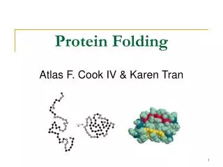
Protein Folding. BL4010 10.10.06. Protein folding is the translation of primary sequence information into secondary, tertiary and quaternary structural information

E N D
Protein Folding BL4010 10.10.06
Protein folding is the translation of primary sequence information into secondary, tertiary and quaternary structural information • Don’t forget post-translational modifications. They change the chemical nature of the primary sequence and thus affect the final structure
other mutants... if vesicle targeting incorrect the proteins are secreted by default
Michel Van Lun http://xray.bmc.uu.se/~michiel/research_files/ext1_10ns.mpg Paul Berg (protein synthesis represented by dance) http://video.google.com/videoplay?docid=-2657697036715872139
Protein Folding Models • Hydrophobic collapse (oil drop model) • entropy driven, temperature dependent • size? • Nucleation theory • alpha helical and reverse turn • Viscosity/collision • Energy landscape
Folded intermediates "Molten globule" When does a protein need help folding?
Protein Folding Machinery • When does protein folding need help? • During synthesis • Upon stress (esp. heat shock) • Players • Chaperone and chaperonins (heat shock factors) • Peptide binding proteins • ATP • Disulfide isomerase • Petidyl prolyl cis-trans isomerase
Improperly folded proteins • Non-functional • improper interactions • nonproductive interactions • Aggregation • Targeted for proteolysis/degradation • Resource drain (commitment of material and energy)
Disulfide isomerization (PDI) • Protein disulfide isomerase • Human protein databank http://www.hprd.org/protein/07181 Cysteine residues can be "tagged" with iodoacetate
Prolyl isomerase • Pin1 catalytic Lys residue "pulls" on the backbone carbonyl
Pin1 an important PPI • Breast cancer: • Pin1 isomerizes phosphorylated Ser/Thr-Pro • Regulates various cellular processes. • Overexpressed in human breast cancer. • Overexpression causes upregulation of cyclin D1 and transformation of breast epithelial cells. • Alzheimer's Disease • Pin1 becomes depleted from the nucleus of diseased neurons • Redirected to the large amounts of abnormally phosphorylated tau proteins that will aggregate into filaments. • This depletion from the nucleus may ultimately contribute to neuronal cell death by reactivating the cell cycle.
What property do misfolded proteins share? • We have seen that protein/protein interactions can be highly specific • Chaperones and Chaperonins must bind a wide variety of different proteins • What is the mechanism of recognition that protein is misfolded?
DnaJ (Hsp40) • Binds folding and misfolded proteins • Contains a 30aa glycine-rich region ('G' domain'), a central Zn binding domain containing 4 repeats of a CXXCXGXG ('CRR' domain) and a C-terminal region of 120 to 170 residues • Associates with DnaK • Discovered as requirement for viral replication
DnaJ hydrophobic = yellow charged = blue
DnaK (Hsp70) • Binds folding and misfolded proteins • Associates with DnaJ • ATP binding and hydrolysis • ER homologue Grp78 • The interaction of DnaK-ATP to substrate proteins is transient and characterized by high on/off rates • 2 domains • alpha helix • beta sheet:peptide binding
dnaJ, dnaK, grpE (foldosome) • Cytoplasmic (ER homologues) • Interaction between the J-domain of DnaJ and the N-terminal ATPase domain of DnaK stimulates ATP hydrolysis. • A second interaction mediated by zinc center II locks DnaK on substrate. • The nucleotide exchange factor GrpE then exchanges ADP with ATP and unlocks DnaK.
Chaperonin complex GroES/GroEL Journal of Biochemistry 2005 137(5):543-549
GroEL (hsp60) Chaperonin • 7-fold symmetry • two rings
GroEl/GroES complex • Free GroEL binds denatured proteins very tightly. • GroEL undergoes an allosteric transition from its tight-binding state to a weaker binding state on the cooperative binding of nucleotides (ATP/ADP) and GroES. • GroES modulates binding of GroEL • 7 ATP molecules bind to GroEL • ATP hydrolysis drives transition back to tight binding state
Folding cage/Iterative annealing • Denatured proteins bind to the resting GroEL·GroES·nucleotide complex. • Fast-folding proteins are ejected as native structures before ATP hydrolysis. • Slow-folding proteins enter chaperoning cycles of annealing and folding after the initial ATP hydrolysis. • Hydrolysis causes transient release of GroES and formation of the GroEL·denatured-protein complexes with higher annealing potential. • The intermediately fast-folding complex is formed on subsequent rebinding of GroES. • The ATPase activity of GroEL·GroES is thus the gatekeeper that selects for initial entry of slow-folding proteins to the chaperone action and then pumps successive transitions from the faster-folding R-states to the tighter-binding/stronger annealing T-states.
Percolator model What happens when an Anfinsen-cage chaperone pulls its target? • The initial volume of the cis cavity (assumed to be 85000 A3) • Water excluded from chamber on protein target • When ATP binds to the cis ring and induces a large conformational change increasing the cavity volume to 175000 A3 • Water moves from the outside (organized structure) to the inside of chaperone and inside of target protein • Influx of water - resolvation of protein • 10 to 15 kcal/mol - provided by the conformational changes driven by ATP-hydrolysis?
Hsp90 • Abundant 2-3% eukaryotic cytoplasmic protein (also nuclear) • ER homologue grp94 glycoprotein folding • ATP binding and hydrolysis in both N- and C-terminal domains • Required for protein folding • Prevents protein aggregates • Deletion embryo lethal ADP Hsp90 N-terminal domain
Calnexin/Calreticulin (lectin chaperones) • Calnexin (CNX)/calreticulin (CNR) are ER chaperones and bind glycoproteins • No homologues in other compartments/Unique action • Calcium binding • CNX membrane anchored/CNR lumenal
Calnexin/Calreticulin • Repeated cycles of deglycosylation, chaperone binding and reglycosylation • Glucosyl transferase is sensor for incomplete folding • Removal of glucose on N-glycans is signal for correct folding/exit to Golgi • Removal of terminal mannose of middle branch of N-glycans is signal for folding failure. Mannose trimming of ER client glycans render them prone to degradation.
Osteosarcoma line Blue Hoescht (DNA) Red Cy3 anti CNX antibody (CNX/ER) Green Phalloidin (Actin cystoskeleton)
CNX/CRT in Immune response • Calnexin binds with partially folded immunoglobulin proteins and retains them in the E.R. until they are completely folded • Recognizes partially glysosylated MHCI • Calreticulin retains MHCI until it binds peptides for display