Download
1 / 25
270 likes | 477 Views
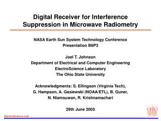
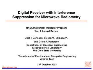
Microwave Radiometry. RTM – Principles. The RTM-01-RES radiometer receives and evaluates the natural electromagnetic radiation (temperature) from the patient’s internal tissues at microwave frequencies. Temperature Distribution. Temperature ( o C). carcinomas of upper outer quadrant.

E N D
Microwave Radiometry
RTM – Principles The RTM-01-RES radiometer receives and evaluates the natural electromagnetic radiation (temperature) from the patient’s internal tissues at microwave frequencies
Temperature (oC) carcinomas of upper outer quadrant carcinomas of inner quadrants 37 36 35 34 33 32 a b d e f g h i j k c 31 Venous and ArterialBlood Temperature
Growth Rate and Specific Heat Production of Breast Carcinomas 70 60 50 40 30 20 10 0 200 100 300 400 500 600 700 Metabolic Heat Production ofCancer Tissueq* (10-3 W/cm3) 128 Breast Carcinomas T1 and T2 Doubling Time of Tumor Volume (days)
Antennas` Field Dx Z
RTM - Diagnosis The efficacy of the method is confirmed by clinical trials, which were carried out among more then 3500 patients
Breast Temperature Field Results of the measurements can be displayed in different modes
RTM-Diagnosis Software Data of the measured temperatures are automatically stored in the computers memory to be processed.
Breast Temperature Field Isotherm step Right MG Left MG Temperature(ºC): Minimum 32.8, Mean 33.8, Maximum 35.3 • The green color represents the average temperature field of an examined organ
Breast Temperature Field Isotherm step Right MG Left MG Temperature(ºC): Minimum 32.8, Mean 33.8, Maximum 35.3 The red color represents a high temperature field of an examined organ
Breast Temperature Field Isotherm step Right MG Left MG Temperature(ºC): Minimum 32.8, Mean 33.8, Maximum 35.3 • The blue color represents a lower than average normal temperature
Healthy Woman’s Field Isotherm step Right MG Left MG Temperature(ºC): Minimum 34.8, Mean 35.1, Maximum 35.4 Example: This is the temperature field of a healthy woman’s breasts.
Breast Temperature Field Isotherm step Right MG Left MG Temperature(ºC): Minimum 32.8, Mean 33.8, Maximum 35.3 Many diseases (such as cancer) are represented by high temperature areas in the internal temperature field.
Sign B Sign C Sign D Sign A Sign A Sign A Sign E Sign F Sign G Sign A Sign A Sign A Expert System The software analyses whether an examined patient has features of cancer.
Thermogram Thermogram - Diagnosis Conventional signs right Mg left Mg base point T1 base point T1 Series 1 Series 2 Series 3 Show all series Print… Help Close The results of the measurements may also be displayed as a thermogram. The fourth and the fifth points on the left breast are marked.
Right Breast Left Breast Isotherm step Conventional signs right Mg left Mg base point T1 base point T2 Series 1 Series 2 Series 3 Show all series Print… Help Close Sign D Sign C Sign B Sign A Sign A Close object: Mark 1 Mark 2 Sign E Sign F Sign G Sign A Sign A Sign A In this case there is an extremely high possibility that the examined patient has left breast cancer
Patient B. (before treatment). Patient B. (after treatment). Right Breast Left Breast Right Breast Left Breast • The RTM-01-RES is indispensable for the monitoring of treatment due to the fact that it is absolutely harmless. • This is a sample of the positive dynamics which occur during the treatment of mastitis.
During oncological processes thermal changes precede anatomical changes. The diagnostic method has a unique ability to detect cancer diseases at early stages, when the traditional methods can not detect diseases, which have just appeared. Patient K., 57 years old Clinical diagnosis - both breasts have diffuse fibrocystic mastopathy, node neoplasm is not detected. X-ray - fibrocystic mastopathy with fibrous features. Puncture - erythrocytes, fat drops.
Clinical Trials Results Number of Patients – 81 CancerPatients – 48 (60%) Number of Patients – 43 CancerPatients – 35 (76%) Number of Patients – 771 CancerPatients – 101 (13%) Number of Patients – 81 CancerPatients – 48 (60%)