Download
1 / 12
120 likes | 281 Views
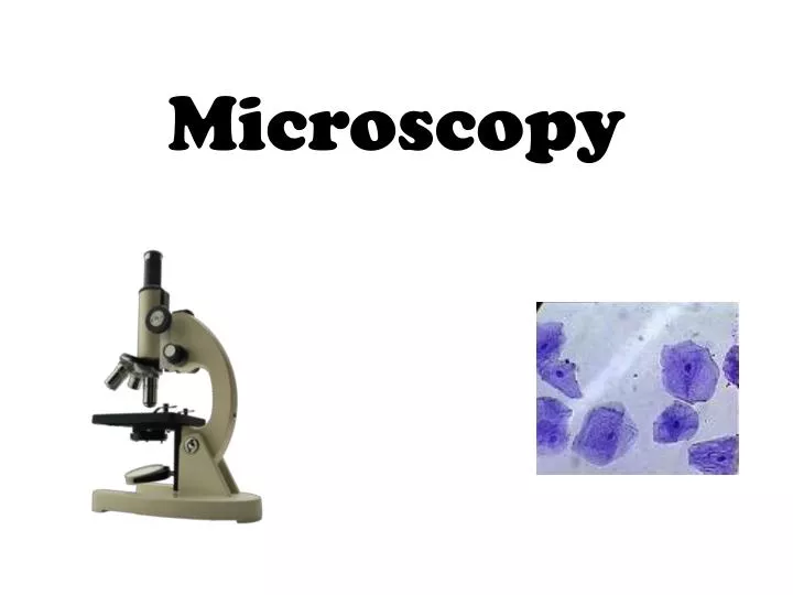
Microscopy. Microscopy Techniques. objective. light source. “transmitted”. Light microscope - transmitting. Microscopy Techniques. light source/objective. reflected dissecting microscope. Stereo or dissecting microscope. Microscopy Techniques. light source/objective.

E N D
Microscopy Techniques objective light source “transmitted”
Microscopy Techniques light source/objective reflected dissecting microscope
Microscopy Techniques light source/objective incident/epi-illumination fluorescent
Fluorescence • fluorescence = process of excitation, loss of energy & emission of light from a fluorophore
Contrast • contrast = difference in color & light between parts of an object/image • required to see an object by microscope • can come from variations in • Intensity (DIC, Phase) • color (bright field, fluorescence) • http://micro.magnet.fsu.edu/primer/techniques/dic/dicphasecomparison.html
Contrast • cells typically are • transparent (not amplitude objects) • phase objects • low in contrast • contrast-generating techniques turn phase differences into intensity differences so we can see unstained cells using transmitted light • DIC (Differential Interference Contrast) microscopy • http://learn.hamamatsu.com/galleries/digitalvideo/spinningdisk/folu473laser/FoLu-EGFP-EB3-100x-1s.html
In Figure 1(c), a thick section of murine kidney tissue imaged with differential interference contrast reveals a bundle of cells enclosed within a tubule. A phase contrast image (Figure 1(d)) of the same area is confusing and disturbed by the presence of phase halos outside the plane of focus.
A human buccal mucosa epithelial (cheek) cell, revealing the nucleus, cytoplasmic inclusions, and numerous bacteria on the upper surface, is presented in Figure 1(a), imaged with differential interference contrast. The same viewfield with phase contrast illumination is illustrated in Figure 1(b). The phase contrast image features pronounced halos around the cellular periphery and nucleus, which are absent in the differential interference contrast image. • Relatively high magnification views of an Obeliapolypoid annulated stem perisarc in DIC and phase contrast are illustrated in Figures 1(e) and 1(f), respectively. In DIC, (Figure 1(e)) the annular structure appears hemispherical with internal rays that emanate radially from the stem. In addition, granular particles are visible within the stem structure, but anatomical detail is largely undefined. The phase contrast image (Figure 1(f)) is confused by halos around the annular rings and within the stem.