Download

1 / 48
600 likes | 1.36k Views
Mechanical Ventilation. Ms. M.N Priyadarshanie. Mechanical Ventilation is ventilation of the lungs by artificial means usually by a ventilator. A ventilator delivers gas to the lungs with either negative or positive pressure. Purposes:.

E N D
Mechanical Ventilation Ms. M.N Priyadarshanie
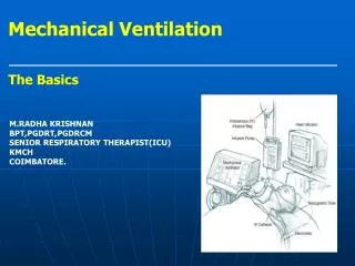
Mechanical Ventilation is ventilation of the lungs by artificial means usually by a ventilator. • A ventilator delivers gas to the lungs with either negative or positive pressure.
Purposes: • To maintain or improve ventilation, & tissue oxygenation. • To decrease the work of breathing & improve patient’s comfort.
Indications: 1- Acute respiratory failure due to: • Mechanical failure, includes neuromuscular diseases as Myasthenia Gravis, Guillain-Barré Syndrome, and Poliomyelitis (failure of the normal respiratory neuromuscular system) • Musculoskeletal abnormalities, such as chest wall trauma (flail chest) • Infectious diseases of the lung such as pneumonia, tuberculosis.
2- Abnormalities of pulmonary gas exchange as in: • Obstructive lung disease in the form of asthma, chronic bronchitis or emphysema. • Conditions such as pulmonary edema, atelectasis, pulmonary fibrosis. • Patients who has received general anesthesia as well as post cardiac arrest patients often require ventilatory support until they have recovered from the effects of the anesthesia or the insult of an arrest.
Types of Mechanical ventilators: • Negative-pressure ventilators • Positive-pressure ventilators.
Positive-pressure ventilators • Positive-pressure ventilators deliver gas to the patient under positive-pressure, during the inspiratory phase.
Classification of positive-pressure ventilators: • Ventilators are classified according to how the inspiratory phase ends. The factor which terminates the inspiratory cycle reflects the machine type. • They are classified as: 1- Pressure cycled ventilator 2- Volume cycled ventilator 3- Time cycled ventilator
Common Ventilator Settings parameters/ controls • Fraction of inspired oxygen (FIO2) • Tidal Volume (VT) • Peak Flow/ Flow Rate • Respiratory Rate/ Breath Rate / Frequency ( F) • I:E Ratio(Inspiration to Expiration Ratio) • Sigh
Signs and symptoms of oxygen toxicity :- 1- Flushed face 2- Dry cough 3- Dyspnea 4- Chest pain 5- Tightness of chest 6- Sore throat
● Pressure Limit • On volume-cycled ventilators, the pressure limit dial limits the highest pressure allowed in the ventilator circuit. • Once the high pressure limit is reached, inspiration is terminated. • Therefore, if the pressure limit is being constantly reached, the designated tidal volume is not being delivered to the patient.
● Sensitivity(trigger Sensitivity) • The sensitivity function controls the amount of patient effort needed to initiate an inspiration • Increasing the sensitivity (requiring less negative force) decreases the amount of work the patient must do to initiate a ventilator breath. • Decreasing the sensitivity increases the amount of negative pressure that the patient needs to initiate inspiration and increases the work of breathing.
Ensuring humidification and thermoregulation • All air delivered by the ventilator passes through the water in the humidifier, where it is warmed and saturated. • Humidifier temperatures should be kept close to body temperature 35 ºC- 37ºC. • In some rare instances (severe hypothermia), the air temperatures can be increased. • The humidifier should be checked for adequate water levels
Ventilator alarms:- • Mechanical ventilators comprise audible and visual alarm systems, which act as immediate warning signals to altered ventilation. • Alarm systems can be categorized according to volume and pressure (high and low). • High-pressure alarms warn of rising pressures. • Low-pressure alarms warn of disconnection of the patient from the ventilator or circuit leaks.
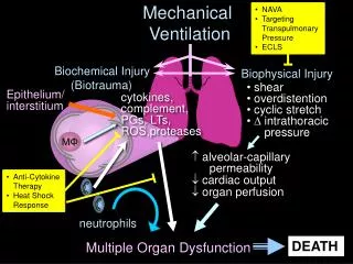
Complications of Mechanical Ventilation:- I- Airway Complications, II- Mechanical complications, III- Physiological Complications, IV- Artificial Airway Complications.
I- Airway Complications 1- Aspiration 2- Decreased clearance of secretions 3- Nosocomial or ventilator-acquired pneumonia
II- Mechanical complications 1- Hypoventilation with atelectasis with respiratory acidosis or hypoxemia. 2- Hyperventilation with hypocapnia and respiratory alkalosis 3- Barotrauma a- Closed pneumothorax, b- Tension pneumothorax, c- Pneumomediastinum, d- Subcutaneous emphysema. 4- Alarm “turned off” 5- Failure of alarms or ventilator 6- Inadequate nebulization or humidification 7- Overheated inspired air, resulting in hyperthermia
III- Physiological Complications 1- Fluid overload with humidified air and sodium chloride (NaCl) retention 2- Depressed cardiac function and hypotension 3- Stress ulcers 4- Paralytic ileus 5- Gastric distension 6- Starvation 7- Dyssynchronous breathing pattern
IV- Artificial Airway ComplicationsA- Complications related toEndotracheal Tube:- 1- Tube kinked or plugged 2- Rupture of piriform sinus 3- Tracheal stenosis or tracheomalacia 4- Mainstem intubation with contralateral (located on or affecting the opposite side of the lung) lung atelectasis 5- Cuff failure 6- Sinusitis 7- Laryngeal edema
B- Complications related to Tracheostomy tube:- 1- Acute hemorrhage at the site 2- Air embolism 3- Aspiration 4- Tracheal stenosis 5- Failure of the tracheostomy cuff 6- Laryngeal nerve damage 7- Obstruction of tracheostomy tube 8- Pneumothorax 9- Subcutaneous and mediastinal emphysema 10- Swallowing dysfunction 11- Tracheoesophageal fistula 12- Infection 13- Accidental decannulation with loss of airway
Nursing care of patients on mechanical ventilation Assessment: 1- Assess the patient 2- Assess the artificial airway (tracheostomy or endotracheal tube) 3- Assess the ventilator
Nursing Interventions 1-Maintain airway patency & oxygenation 2- Promote comfort 3- Maintain fluid & electrolytes balance 4- Maintain nutritional state 5- Maintain urinary & bowel elimination 6- Maintain eye , mouth and cleanliness and integrity:- 7- Maintain mobility/ musculoskeletal function:-
Nursing Interventions 8- Maintain safety:- 9- Provide psychological support 10- Facilitate communication 11- Provide psychological support & information to family 12- Responding to ventilator alarms /Troublshooting ventilator alarms 13- Prevent nosocomial infection 14- Documentation
Responding To Alarms • If an alarm sounds, respond immediately because the problem could be serious. • Assess the patient first, while you silence the alarm. • If you can not quickly identify the problem, take the patient off the ventilator and ventilate him with a resuscitation bag connected to oxygen source until the physician arrives. • A nurse or respiratory therapist must respond to every ventilator alarm.
Alarms must never be ignored or disarmed. • Ventilator malfunction is a potentially serious problem. • Nursing or respiratory therapists perform ventilator checks every 2 to 4 hours, and • Recurrent alarms may alert the clinician to the possibility of an equipment-related issue.
Causes of Ventilator Alarms High pressure alarm • Increased secretions • Kinked ventilator tubing or endotracheal tube (ETT) • Patient biting the ETT • Water in the ventilator tubing. • ETT advanced into right mainstem bronchus.
Low pressure alarm • Disconnected tubing • A cuff leak • A hole in the tubing (ETT or ventilator tubing) • A leak in the humidifier
Oxygen alarm • The oxygen supply is insufficient or is not properly connected.
High respiratory rate alarm • Episodes of tachypnea, • Anxiety, • Pain, • Hypoxia, • Fever. -
Apnea alarm • During weaning, indicates that the patient has a slow Respiratory rate and a period of apnea.
Temperature alarm • Overheating due to too low or no gas flow. • Improper water levels
Methods of Weaning 1- T-piece trial, 2- Continuous Positive Airway Pressure (CPAP) weaning, 3- Synchronized Intermittent Mandatory Ventilation (SIMV) weaning, 4- Pressure Support Ventilation (PSV) weaning.
Weaning readiness Criteria • Awake and alert • Hemodynamically stable, adequately resuscitated, and not requiring vasoactive support • Arterial blood gases (ABGs) normalized or at patient’s baseline - PaCO2 acceptable - PH of 7.35 – 7.45 - PaO2 > 60 mm Hg , - SaO2 >92% - FIO2 ≤40%
Positive end-expiratory pressure (PEEP) ≤5 cm H2O • F < 25 / minute • Vt 5 ml / kg • VE 5- 10 L/m (f x Vt) • VC > 10- 15 ml / kg • PEP (positive expiratory pressure) > - 20 cm H2O ( indicates patient’s ability to take a deep breath & cough),
Chest x-ray reviewed for correctable factors; treated as indicated, • Major electrolytes within normal range, • Hematocrit >25%, • Core temperature >36°C and <39°C, • Adequate management of pain/anxiety/agitation, • Adequate analgesia/ sedation (record scores on flow sheet), • No residual neuromuscular blockade.
Role of nurse before weaning:- 1- Ensure that indications for the implementation of Mechanical ventilation have improved 2- Ensure that all factors that may interfere with successful weaning are corrected:- - Acid-base abnormalitie - Fluid imbalance - Electrolyte abnormalities - Infection - Fever - Anemia - Hyperglycemia - Protein - Sleep deprivation
Role of nurse before weaning:- 3- Assess readiness for weaning 4- Ensure that the weaning criteria / parameters are met. 5- Explain the process of weaning to the patient and offer reassurance to the patient.
6- Initiate weaning in the morning when the patient is rested. 7- Elevate the head of the bed & Place the patient upright 8- Ensure a patent airway and suction if necessary before a weaning trial, 9- Provide for rest period on ventilator for 15 – 20 minutes after suctioning.
10- Ensure patient’s comfort & administer pharmacological agents for comfort, such as bronchodilators or sedatives as indicated. 11- Help the patient through some of the discomfort and apprehension. 12- Support and reassurance help the patient through the discomfort and apprehension as remains with the patient after initiation of the weaning process. 13- Evaluate and document the patient’s response to weaning.
Role of nurse during weaning:- 1- Wean only during the day. 2- Remain with the patient during initiation of weaning. 3- Instruct the patient to relax and breathe normally. 4- Monitor the respiratory rate, vital signs, ABGs, diaphoresis and use of accessory muscles frequently. If signs of fatigue or respiratory distress develop. • Discontinue weaning trials.
Signs of Weaning Intolerance Criteria • Diaphoresis • Dyspnea & Labored respiratory pattern • Increased anxiety ,Restlessness, Decrease in level of consciousness • Dysrhythmia,Increase or decrease in heart rate of > 20 beats /min. or heart rate > 110b/m,Sustained heart rate >20% higher or lower than baseline
Increase or decrease in blood pressure of > 20 mm Hg . Systolic blood pressure >180 mm Hg or <90 mm Hg • Increase in respiratory rate of > 10 above baseline or > 30. Sustained respiratory rate greater than 35 breaths/minute • Tidal volume ≤5 mL/kg, Sustained minute ventilation <200 mL/kg/minute • SaO2 < 90%, PaO2 < 60 mmHg, decrease in PH of < 7.35. Increase in PaCO2
Role of nurse after weaning 1- Ensure that extubation criteria are met . 2- Decannulate or extubate 2- Documentation