Download
1 / 32
320 likes | 452 Views
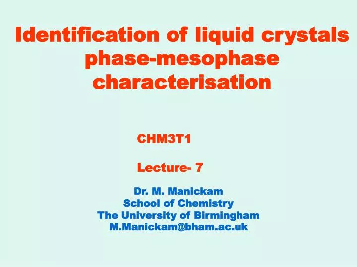
Identification of liquid crystals phase-mesophase characterisation. CHM3T1 Lecture- 7. Dr. M. Manickam School of Chemistry The University of Birmingham M.Manickam@bham.ac.uk. Outline of Lecture. Introduction Thermal Analysis Polarised Optical Microscopy

E N D
Identification of liquid crystals phase-mesophase characterisation CHM3T1 Lecture- 7 Dr. M. Manickam School of Chemistry The University of Birmingham M.Manickam@bham.ac.uk
Outline of Lecture • Introduction • Thermal Analysis • Polarised Optical Microscopy • Differential Scanning Calorimetry • Mesophase Textures • X-Ray diffraction
Learning Objectives After completing this lecture you should have an understanding of, and be able to demonstrate, the following terms, ideas and methods. • Polarised Optical Microscopy (POM) • Reflection and Refraction • Index of Refraction • Birefringence • Mesophase Textures • Differential Scanning Calorimetry (DSC) • X-Ray Diffraction
Examples Example of a compound that shows no LCs phase liquid water 0 degrees of order solid crystalline water; 3- (dimensional) degrees of order gaseous water 0 degrees of order Example of a compound that shows LCs phases Looks like milk 1 degree of order 3 degrees of order 0 degrees of order 0 degrees of order 3 degrees of order in solid form gooey material 2 degrees of order
Thermal Analysis Thermal Methods of Analysis The first step in the investigation of the liquid crystalline nature of materials is based upon thermal methods of analysis. When a mesomorphic material in the crystal state is subjected to heating, the energy supplied disrupts the crystalline lattice leading to the LC phase. As the temperature rises, the LC will absorb further energy becoming an isotropic liquid. Thermal analysis allows the detection of this sequence of the phase transitions, using • Polarised optical microscopy (POM) • Differential scanning calorimetry (DSC)
Polarised Light and Unpolarised Light (a) (b) • Representation of the unpolarised light, travelling in the direction perpendicular to the page. The electric (and magnetic) field vibrates in all the possible planes (represented by the arrows) perpendicular to the propagation of the light. • Polarised light is characterised by only one plane of polarisation of the electric (and magnetic) field, which is represented by the vertical arrow. Polarised light (figure- a & b) is generated by the passage of unpolarised light (white light) through a polariser. The polariser is a transparent anisotropic material, which selectively allows the transmission of light along one preferential plane of polarisation, which corresponds to the polariser optical axis. Examples of such kinds of materials are calcite prisms (e.g. Nicol prism) and polarising Filters (e.g. Polaroid).
Light Travelling in a Vacuum Electromagnetic Radiation In vacuum light travels at 300 X 10 6 ms-1
Light Travelling Through an Isotropic Medium X and Y polarised light travelling through an isotropic medium One Values for n
Light Travelling Through an Anisotropic Medium X and Y polarised light travelling through an anisotropic medium Two Values for n
Index of Refraction ~ Light travelling through a vacuum does so at a velocity of ~ 3 X 10 8 ms-1, however this changes in the presence of matter. The electric and magnetic fields of a light wave affect the charges in a material causing them also to produce electric and magnetic fields. The net effect of this is that the velocity of light passing through matter is less than that passing through a vacuum. This retardation varies with the nature of the material, and each material is assigned a number that represents the factor by which the velocity of light is reduced. This is called the index of refraction, n, and is defined as: n = c / v Where: c = the velocity of light in a vacuum v = the velocity of light in a material
Index of Refraction The index of refraction of all materials is greater than one; the following values are for comparison Indices of refraction for some common materials
Reflection and Refraction of Light at the Surface of an Isotropic Materials
Reflection and Refraction of Light at the Surface of an Anisotropic Materials Birefringence or Double Refraction
Polarised Optical Microscopy (POM) POM is employed to observe the mesophase textures of LCs, exploiting their anisotropic nature and, in particular, their birefringence when interacting with polarised light. Polarised optical microscopes are equipped with two polarisers (a polariser and an analyser), whose relative optical axis can be rotated from 0o to 900, changing from a parallel to a perpendicular arrangement respectively. If the two polarisers are set up in series (at 0o) their optical axes are parallel, consequently light passes through both (figure a). When they are in a crossed position (at 90o), their axes are perpendicular, therefore light from the first is extinguished by the second (figure b). In order to investigate the mesophase behaviour of LCs, the most commonly informative and used setting for the two polarisers is the crossed (90o) position.
Polariser and Analyser Figure : (a) When the polariser and analyser are in a parallel set up, their optical axes allow light transmission; (b) when the polariser and analyser are crossed, the light from the polariser is absorbed by the analyser, resulting in dark condition.
Birefringence in LCs When polarised light enters an anisotropic material (e.g. LC) it splits into two components, the ordinary and extraordinary rays, whose electric (and magnetic) fields vibrate in fixed planes at right angle to each other and propagate through the material at different velocities. As a consequence of the delay of one ray over the other, the two waves become out of phase. Therefore, the plane of polarisation of the light is rotated. Thus, when the polarised light reaches the analyser, there will be a component of it, which can go through its optical axis, and the light will be transmitted. The preferential orientation of the molecules along the director, which forms an angle other than 0o or 90o with either the polariser or the analyser, is responsible for the rotation of the plane of polarisation and transmission of light with production of a bright field of view. Hence, when a LC is placed between two crossed polarisers, it will shine bright interference colours, giving a characteristic pattern, which represents the “finger- print” texture of the mesophase.
Birefringence Birefringence is the term applied to the double refraction of nonpolarised light as it passes through an anisotropic material. This phenomenon occurs because the x-polarised and y- polarised component of the light interact differently with the anisotropic material, giving rise to two refractive indices, and therefore two refracted light beams, as illustrated in the figure.
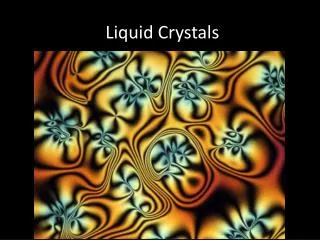
Polarized microscopy of the mesophases Examples of OPM images R= C5H11 R= C5H11 Optical texture of Ester at 70 0C Optical texture of Ether at 50 0C
Mesophase textures Schlieren texture of Nematic Fan-shaped texture of smectic
Mesophase Textures batonnets smectic Focal conic textures of smectic
Mesophase Texture B2 B1 Banana-shaped LC
Mesophase Textures B4 phase B3 phase Banana-shaped LC
Differential Scanning Calorimetry (DSC) Whenever a material undergoes a change in physical state, heat (Q) is either absorbed (e.g. melting) or liberated (e.g. solidification). By monitoring calorimetrimetrically, the temperature change (ΔT) that accompanies a phase transition, it is possible to measure the energy involved, as a variation of enthalpy (ΔH), which is typical of the material for the transition under study. Therefore, useful information for the characterisation of compounds is obtained by the calculation of ΔH. DSC is one of the most widely used sophisticated methods to investigate samples behaviour over a range of programmed temperatures at constant pressure. The term “differential scanning calorimetry” summarises the nature of the thermal technique involved.
Differential Scanning Calorimetry (DSC) Calorimetry: the sample and an inert reference (commonly dry pre-heated alumina) are heated, simultaneously, at a defined rate, in an inert atmosphere at constant pressure over a programmed range of temperature Scanning: the temperature of the system is scanned over a desired range as a function of time. Differential: the difference in heat flow or power, ΔP (ΔP = dΔQ / dt) required to maintain the sample and the reference at the same temperature, is measured and plotted against temperature or time in a x y graph (since the thermal analysis is run under constant pressure, the measure of the heat corresponds to the enthalpy: ΔQ = Δ H). An endotherm peak (ΔH< 0) is involved when there is absorption of more power by the material under analysis respect to the reference, whilst an exothermic peak (ΔH > 0) underlines absorption of more power by the reference, implying a liberation of energy by the analyzed material. Plotting of the peaks upward or downward is a matter of convention.
Differential Scanning Calorimetry (DSC) Figure-a Figure-a: DSC trace showing the typical pattern of a LC exhibiting a crystal to mesophase (K M) transition at 65.8oC, and a mesophase to isotropic liquid (MI) transition at 95.7oC. The endothermic peaks go up, and exothermic ones go down: y, heat flow (mW); x, temperature (oC)
Differential scanning calorimetry (DSC) From the DSC analysis it is possible to obtain the following quantitative data: • T: onset temperature of phase transition (by differentiation), • As: peaks area (by integration), • Δ H: enthalpy change of phase transition (by integration). The measurement of Δ H is very useful to determine the entropy change (ΔS) associated with physical changes of LCs. In fact at a transition temperature, any exchange of heat between the sample and the surrounding is reversible, because the two phase are in equilibrium. Therefore, it is possible to calculate the change in entropy (Δ S = Δ H/ T).
DSC Apparatus The major parts of the system: 1. the DSC sensors plus amplifier, 2. the furnace and its temperature sensor, 3. the programmer or computer, 4. the recorder, plotter or data acquisition device Δ indicates the differential signal
DCS: B7 phase of Banana-Shaped Achiral Mesogen The DSC thermogram obtained using heating and cooling modes (5 oC min-1) is shown in Figure Only one mesophase is observed in both cyles. 112.7oC 172.7oC
DSC Thermograms fo Anthraquinone-based Discotic The DSC runs were recorded at a heating / cooling rate of 5 oC min-1 77.0 143.5 128.9 I Cr Colx transition Colh 113.6 Colh 140.0 I 1,5-benzloxy-2,3,6,7- Tetraalkyloxy-9,10- anthraquinones 127.7 Colh 143.5 I DSC thermograms for (i) the first heating; (ii) second heating, and (iii) first cooling
X-Ray diffraction studies Intensity (arb.units) 10 20 30 0 2(deg) R= C5H11 R= C5H11 intracolumnar intracolumnar - alkyl Intensity (arb.units) - alkyl 0 10 20 30 2(deg) The overall features observed are consistent with the structure of the Colhphase
Bragg Equation When a beam of monochromatic X-rays of wavelength λ impinges on a crystal, strong scattering occurs in certain directions only: this is the phenomenon of X- Ray Diffraction nλ = 2d sinθ n= (1, 2, 3……., ) wavelengths d= is the distance separating successive planes in the crystal θ = is the angle which the incident beam X-rays makes with the same planes
Final Comments Identification and systematic classification of scientication of scientific phenomena is vital in any area of research. Liquid crystals are no exception and many different liquid crystalline phases and other mesophases have been identified and classified according to their distinct phase structures. Many liquid crystal phases (e.g., nematic, smectic A, smectic C and their chiral analogues) are commonly encountered in a wide range of compounds of varying molecular architectures. Such liquid crystal phases are now easily identified by using optical polarising microscopy, usually in conjunction with differential scanning calorimetry. However, some liquid crystal phases (e.g., antiferroelectric and ferrielectric phases ) are relatively recent discoveries and are more rarely encountered. Although such novel LC phases can usually be identified by optical microscopy, their phase structures have not yet been fully elucidated and so other techniques such as X-ray analysis must be used. Accordingly, just as the field of liquid crystals draws on the expertise of scientists from many disciplines, the identification of mesophases requires a wide range of techniques to identify and classify fully the different structures of the various mesophases. As the identification techniques become more sophisticated, more novel mesophases will be discovered, possibly paving the way for the development of more technological applications.