Download
1 / 84
1.04k likes | 1.52k Views
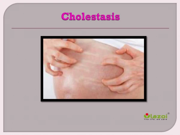
Management Of Neonatal Cholestasis. Dr. Shrish Bhatnagar Consultant Paediatric Gastroenterologist Eras’ Lucknow Medical College & Vivekananda Polyclinic and Institute of Medical Sciences, Lucknow. TOPICS. Definition Incidence & Etiology Bile Acid Metabolism Classification

E N D
Management Of Neonatal Cholestasis Dr. ShrishBhatnagar Consultant Paediatric Gastroenterologist Eras’ Lucknow Medical College & Vivekananda Polyclinic and Institute of Medical Sciences, Lucknow.
TOPICS • Definition • Incidence & Etiology • Bile Acid Metabolism • Classification • Clinical features • Investigations • Treatment • Specific Disease • Summary
DEFINITION • Neonatal Cholestasisis defined biochemically as prolonged elevation of the serum levels of conjugated bilirubinbeyond the first 14 days of life. (1) (NELSON textbook of pediatrics)
Conjugated hyperbilirubinemia is generally defined as a conjugated or direct bilirubin level greater than 1 mg/dL when the total bilirubin is less than 5 mg/dL or more than 20% of the total bilirubin if the total bilirubin is greater than 5 mg/dL. • Conjugated hyperbilirubinemia is never physiologic or normal. • The preferred term to describe it is cholestasis, which includes retention of CB ,bile acids, cholesterol and other components of bile.(2) JOHN P. CLOHERTY
The incidence of neonatal cholestasis is around 1 in 2500 live births in the west.(3) • In India it constitutes 30% of all hepatobiliary disorders.(4)
ETIOLOGY • 1. LIVER CELL INJURY-(normal bile ducts) A. Toxic B. Infection C.Metabolic Viral Bacterial hepatitis (B,C) syphilis, E.coli Rubella, AdenoListeria, tuberculosis Herpes, Ebv staphylococcus Echovirus 14 β-hemolytic streptococcus C. Metabolic - α 1-antitrypsin def., cystic fibrosis , galactosemiaTyrosenemia, Fructosemia, storage diseases (gaucher , niemann-pick, glycogenosis type 4,dubin-johnson syndrome, byler disease, zellweger syndrome, idiopathic cirrhosis, porphyria,
ETIOLOGY 2.Bile flow obstruction Biliaryatresia : Extrahepatic- Choledochal cyst, Trisomy 13 /18 Intrahepatic- PILBD ( Syndromic :Alagille syndrome & Non Syndromic ) Intrahepaticatresia with lymphedema (Aagenaes syndrome),PFIC, Rupture of bile duct , Pancreatic cyst , Hemangiomas , Tumors , Inspissated bile syndrome.
Global Data 12 counties/ 5 continents
Classification Nelson textbook of pediatrics Neonatalcholestasis Extrahepatic disease (bile duct injury /obstruction) Intrahepatic disease Bile duct injury Hepatocyte injury Biliaryatresia PFIC/PSC PILBD Metabolic ds. Viral ds.
Working algorithm for management of neonatal cholestasis NC Sick (7) Not sick Sepsis screen +ve Sepsis screen +ve Pigmented stools Pale stools Treat sepsis (galactosemia needs to be excluded by urinereducing sugar ) Test for CMV, HSv urinary succinylacetone ,ferritin, GALT if urine reducing sugar positive Fasting USG (see for contractility post feed) GGT ,fasting USG,metabolic test , genetic test , liver biopsy Specific treatment Normal Small gall bladder Absent gall bladder Choledochal cyst Liver biopsy Biliaryatresia to be excluded surgery Specific treatment
Clinical features • The typical findings in an infant who has cholestasis are protracted jaundice, scleralicterus, acholic stools, dark yellow urine, and hepatomegaly. • Acholic stools in an infant should always prompt further evaluation. • Some infants may have coagulopathy secondary to vitamin K malabsorption and deficiency and present with bleeding or bruising. • Splenomegaly can be observed in infants who have cirrhosis and portal hypertension, storage diseases, and hemolytic disorders.
Clinical features • Neurologic abnormalities including irritability, lethargy, poor feeding, hypotonia, or seizures can indicate sepsis, intracranial hemorrhage, metabolic (like Galactosemia ) • Low birth weight, thrombocytopenia, and chorioretinitis are often associated with congenital infection. • Facial dysmorphism may suggest a chromosomal abnormality or Alagille syndrome. • A palpable mass in the right upper quadrant may indicate a choledochal cyst. • A cardiac murmur increases the likelihood of Alagille syndrome or BA.
Investigations • If cholestasis is present, further evaluation should be completed with a sense of urgency because patients who have BA have a better outcome if they undergo a Kasai hepatic portoenterostomy (HPE) before age 30 to 45 days, • Levels of liver enzymes, including SGOT, SGPT, and ALP , are usually elevated in a cholestatic infant but are poor predictors of etiology. • γ-Glutamyltranspeptidase (GGT) is generally elevated during cholestasis however, • A low or normal GGT progressive familial intrahepaticcholestasis (PFIC) type 1, PFIC type 2, an inborn error of bile acid synthesis or metabolism, or panhypopituitarism. • Baseline albumin, glucose, and prothrombin time/international normalized ratios are useful in assessing the degree of liver synthetic dysfunction.
Investigations • Depending on the clinical scenario, bacterial cultures from blood and urine may be indicated. • Among the congenital infections, cytomegalovirus (CMV) is most commonly implicated. Ig G/M levels are unreliable • Assay of pp65 antigen and CMV polymerase chain reaction (PCR) are more specific and reliable if the biochemical tests and histology are consistent with the diagnosis. • Screening Test • Alpha feto protein High S/o Tyrosenimia • Ur NGRS 2/3 positive S/o Galactosemia • S Ferritin High S/o Neonatal Hemochromotosis • Raised immunoreactivetrypsinogen S/o Cystic fibrosis
Investigations An abdominal USG to assess liver structure, size, and composition; to evaluate for the presence of ascites; and to identify findings of biliaryatresia or an extrahepatic obstructive lesion (choledochal cyst, mass, gallstone, and sludge ). A chest radiograph may reveal cardiomegaly or butterfly vertebrae in patients who have Alagille syndrome
Investigations • Cardiac Murmur: 2 D echocardiogram should be obtained. Up to 24% of patients who have Alagille syndrome have cardiac abnormalities , Rubella (Peripheral Pulmonary Stenosis) • Eye Examination : • Cataract: Galactosemia • Post embryotoxon : Alagille syndrome • Chorio Retinitis : CMV • Cherry red Spot: Niemann Picks and Tay Sachs
TREATMENT • Most infants with NC are underweight and will need nutritional support. • The goal is to provide adequate calories to compensate for steatorrhea and to prevent / treat malnutrition. • The calorie requirement is approximately 125% of the recommended dietary allowance (RDA) based on ideal body weight .(13) • In breastfed infants, breastfeeding should be encouraged and medium-chain triglyceride (MCT) oil should be administered in a dose of 1-2 mL/kg/d in 2-4 divided doses in expressed breast milk . (14)
TREATMENT • In older infants, a milk-cereal-mix fortified with MCT is preferred. • Adding puffed rice powder and MCT to milk can make feeds energy-dense. Essential fatty acids should constitute 2- 3% of the energy provided. Vegetable protein at 2-3 g/kg/d is recommended .(14) • Infants with cholestasis require supplementation with fat-soluble vitamins administered orally as water-soluble preparations.
TREATMENT • In treatment of vitamin deficiencies, standard deficiency protocols should be followed. • 1,25 dihydroxy Vitamin D3 (0.05-0.2ug/kg/d) is recommended in the presence of significant bone changes or patients having severe cholestasis. • Vitamin K is administered at a dose of 5mg intramuscular, subcutaneously or intravenously for 3 days, at diagnosis to correct the coagulopathy.
TREATMENT • If the INR is markedly prolonged, intramuscular injections should be avoided. • Vitamin supplementation should be continued till 3 months after resolution of jaundice. (15)
SUGGESTED DAILY VITAMIN AND MINERAL REQUIREMENTIN INFANTS WITH CHOLESTASIS
SPECIFIC TREAMENT In infants with pruritus due to severe cholestasis, the drug given in the following order: • Ursodeoxycholic acid (UDCA) (20 mg/kg/d), • rifampicin(5-10 mg/kg/d), • phenobarbitone (5–10 mg/kg/d). • Symptom chart should be made for pruritus. • Depending on severity and response to previous agent, add-on drug can be considered. • Appropriate antibiotics depending on the site of infection and culture sensitivity reports need to be administered
Case I: 50 days girl ELMCH @ 50 d FTND, (wt 2.8kg) Admitted for 5 day at birth else where ? Sepsis/ Jaundice ( PT =48hrs) Wt : 4.8 Kg Well Playful Icterus + Liver /Spleen + Few Skin Bleeds ( reason for Referral) H/o Clay Colored Stool Jaundice x 20 days Top fed Bottle feeding Clinical Diagnosis : EHBA
BILIARY ATRESIA • It is an idiopathic inflammatory process involving the bile ducts resulting in obstruction of biliary tract, chronic cholestasis and progressive fibrosis and eventually to biliary cirrhosis.
Perinatal infection Abnormal BD development Gene mutation Perinatalbiliaryatresia Embryonic biliaryatresia Bile duct epithelial cell injury expression of Ag BD obstruction Bile flow initiation Infammatory ?autoimmune response ?Genetic predisposition Hepatocellular injury BD – apoptosis,fibrosis, obstruction ? modifier genes Intrahepaticcholestasis and inflammation Fibrosis and biliary cirrhosis
BILIARY ATRESIA • The most common form of biliaryatresia is obliteration of the entire extrahepaticbiliary tree at or above the portahepatis. • Most patients are normal at birth and have a postnatal pogressive obliteration of bile ducts. • The embryonic form manifest at birth and associated with- situsinversus, polysplenia, intestinal malrotation , complex congenital heart disease.
Anatomically BA is of three types ; 1) involving common bile duct and patent proximal biliary system 2) atresia involving hepatic duct with patent proximal ducts 3) involving right and left hepatic ducts at the portahepatis (most common 85%)
CLINICAL PRESENTATION • If jaundice is associated with dark urine and/ or pale stool, is suggestive of cholestasis. • The sensitivity, specificity and positive predictive value of pale stools for the detection of biliaryatresia (BA) before 60 days as determined by a color-coded stool chart was noted to be 89.7%, 99.9% and 28.6%, respectively.(8)
CLINICAL PRESENTATION • Babies with BA appear well and have normal growth and development in spite of their jaundice, and this leads to parents and physicians underestimating the seriousness of the problem . • Persistent cholestasis from any cause leads to liver damage and cirrhosis. Therefore, determining the specific etiology (medical or surgical) at the earliest is critical.
INVESTIGATIONS • Abdominal ultrasonography findings described in BA include the triangular cord sign, abnormal gallbladder morphology (not visualized or length <1.9 cm or lack of smooth/complete echogenic mucosal lining with an indistinct wall or irregular/lobular contour) , no contraction of gall bladder after oral feeding and nonvisualized common bile duct (CBD). • A distended gall bladder, however, does not rule out a proximal BA with a distal patent bile duct and mucus filled gallbladder. • It is recommended that ultrasound should be done after 4 hours of fasting.
Triangular cord sign: Abnormal triangular echogenic area in the region of the portahepatis ,representing fibrous remnant of hepatic duct.(10)
INVESTIGATIONS • Hepatobiliary-imino-di-acetic acid (HIDA) scan has limited role in evaluation of NC especially if the baby has clearly documented pale or pigmented stools. • The time required (3 days) for priming before the scan, especially in patients who are referred late, is a limitation. • Priming done by UDCA at dose of 20/mg/d
INVESTIGATIONS • Performing a HIDA scan is optional and one may go for a liver biopsy straight away. • HIDA is, however, useful in the diagnosis of the uncommon causes like spontaneous perforation of the bile duct .(11) • Intra-operative cholangiogram (IOC) remains the gold standard for diagnosis of BA. • Liver biopsy is an essential investigation in the evaluation of NC.
HIDA SCAN (Hepatobiliary-imino-di-acetic acid) • A hepatobiliary (HIDA) scan is an imaging procedure used to diagnose problems of the liver, gallbladder and bile ducts. • HIDA scan is also known as cholescintigraphy and hepatobiliaryscintigraphy.
HIDA SCAN • A radioactive tracer is injected through any accessible vein and then allowed to circulate to the liver, where it is excretedinto the bile ducts and stored by the gallbladder until released into the duodenum.
HIDA SCAN • In the absence of gallbladder disease, the gallbladder is visualized within 1 hour of the injection of the radioactive tracer. • If the gallbladder is not visualized within 4 hours after the injection, this indicates either cholecystitis or cystic duct obstruction, such as by cholelithiasis (gallstone formation).
USES OF HIDA SCAN These include: • gallbladder inflammation, or cholecystitis • bile duct blockages • Congenital bile duct abnormalities, such as biliaryatresia • An excretory HIDA rules out BA • A Non excretory HIDA is not diagnostic of BA can be seen in Severe Cholestasis
RESULT OF HIDA SCAN • Normal -The radioactive tracer moved freely with bile from the liver into gallbladder and small intestine. • Slow- The tracer moved slower than normal . This may be a sign of a blockage or a problem with liver. • Not present- If there are no signs of radioactive tracer in gallbladder on the images, this may be a sign of acute gallbladder inflammation, or acute cholecystitis. • Radioactive tracer in other parts of the body- If images show signs of radioactive tracer outside of liver, gallbladder, bile ducts, and small intestine, may have a leak in biliary (bile) system.
HIDA SCAN • The scan is also important to differentiate between neonatal hepatitis and biliaryatresia, because an early surgical intervention in form of Kasai portoenterostomy or hepatoportoenterostomy can save the life of the baby as the chance of a successful operation after 3 months seriously decreases.
LIVER BIOPSY • Liver biopsy is the removal of a small sample of tissue from the liver. It is a medical test that is done • to aid diagnosis of liver disease, • to assess the severity of known liver disease, • to monitor the progress of treatment.
LIVER BIOPSY • Types • Percutaneously (via a needle through the skin), • Transvenously (through the blood vessels), • Endoscopically (through endoscopic ultrasound fine needle biopsy) • Directly during abdominal surgery. The sample is examined by microscope,