Download

1 / 23
230 likes | 364 Views
Comlication of IOL implantation. Dr A.Ashtari Isfahan university of medical sciences. Decentration and Dislocation. Decentration may occure in following situation Asymetric haptic placement,with one haptic in the bag and the other in the sulcus

E N D
Comlication of IOL implantation Dr A.Ashtari Isfahan university of medical sciences
Decentration and Dislocation Decentration may occure in following situation • Asymetrichapticplacement,with one haptic in the bag and the other in the sulcus • Insufficient zonular or capsular support • The presence of irregular fibrosis of the post capsul • Capsular phimosis
Decentration can produce unwanted glare and reflection if the edge of lens is within the pupillary space
If zonular support is inadequate the surgeon should attempt to rotate the IOL to a position where clinical evidence shows sufficient capsule and zonular fibers to support the implant The use of transcorneal iris fixation sutures(Mc-Cannel sutures)to secure the IOL may also be considered
Irregular posterior capsul fibrosis gradually decenters the IOL. Deformation of the haptics may render simple rotation insufficient to center the IOL properly It may become necessary in these cases to move the IOL haptics into the ciliarysulcus or replace the capsul fixated IOL with a posterior chamber sulcus-fixated IOL.
If dislocation of the IOL is complete the surgeon can sublux the optic of the implant into the pupil by means of vitrectomy technique and use transcorneal iris-fixation sutures to fix the 2 haptics of the implant Altenatively,the implant may be removed altogether and replaced with either an anterior chamber IOL or a transscleral or iris-sutured posterior chamber IOL.
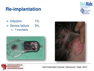
Subluxatio of scleral-fixated sutured IOL has been reported 3-9 years after implantation with 10-0 polypropylene fixation sutures.Double-fixation techniques and thicker 9-0 polypropylene sutures are currently recommended for scleral fixation of IOLs.
Pupillary capture Causes: • Formation of synechiae between the iris and underlying posterior capsule • Improper placement of the IOL haptics • Shallowing of the anterior chamber • Anterior displacement of the posterior chamber IOL optic
Ant displacement of post chamber IOL optic is associated with: • Placement of non angulated IOL in ciliarysulcus • Upside-down placement of an angulated IOL so that the IOL angles anteriorly • Positive vitreous pressure
Placement of a posteriorly angulated post chamber IOL in the capsular bag decrease the likelihood of pupillary capture
Usually,pupillary capture is a purely cosmetic issue;the patient is otherwiseasymptomatic and can be left untreared Occasionally pupillary capture can cause problem such as: • Glare • Photophobia • Chronic uveitis • Unintended myopia • Monocular diplopia
Mydriatic can sometimes be used succesfully to free the iris through pharmacologic manipulation of the pupil If conservative management fails,surgical intervention may be required to free the iris,break the synechiae,or reposition the lens
Capsular block syndrom Capsular block syndrome is an uncommon postoperative complication of capsular bag-fixated posterior chamber IOL Aqueous becomes trapped within the capsular bag,between the post capsul and the post surface of the IOL There is forward displacement of the lens optic with a resultant myopic shift The fluid behind the IOL may have a turbid or milky appearance
Nd:YAG laser post capsulotomy results in release of the fluid,post movement of the IOL optic to its original position,and resolution of the myopic shift
Uveitis-glaucoma-hyphemasyndrom First described in the context of rigid ant chamber and closed-loop IOLs The classic triad or individual elements may occure as a result of inappropriate IOL sizing,contact between the implant and vascular structure or the corneal endotheliumor defect in implant manufacturing
UGH can also occure in pt with post chamber IOL owing to contact between the lens loops and uvealtissuein the post chamber UGH may respond to treatment with topical anti inflamatory medication or anti glaucoma medication If the symptoms are not alleviated sufficiently by mediacl therapy or inflammation threatens either retinal or corneal function,IOL removal must be considered
This procedure may be very complicated because of inflammatory scars,particulary in the angle If such scarring is present,the surgeon may need to amputate the haptics from the optic and remove the lens piecemeal,rotating the haptics material out of the synechial tunnels to minimize trauma to the eye. In some cases it is safer to leave portions of the haptics in places Early lens explantation may reduce the risk of corneal decompensationand CME
Pseudophakicbullouskeratopathy Certain IOL design,particularly iris-clip lenses(iris-fixated lenseswith the optic anterior to the iris)and closed-loop flexible anterior chamber lenses,are associated with increased risk of corneal decompensation Iris –clip lenses have been shown to contact the corneal endothelium during eye movement Chronic endothelial cell loss associated with closed-loop IOL is thought to be due to chronic inflammation and contact between the lens and peripheral corneal endothelial cells Both types of lenses are no longer in clinical use
Patinets with underlying corneal dysfunction such as Fuchs dystrophy are at greater risk fer developing postoperative corneal edema. • Progressive stromal edema eventually leads to bullouskeratopathy
IolDesign,Glare,andOpacification In addition to lens decentration and capsular opacification,glare can result when the diameter of the IOL optic is smaller than the diameter of the scotopic pupil Optics with a square-edge design and multifocal IOL are more prone to producing glare and halo Spherical aberration may produce some degree of distortion or glare under scotopic conditions when the pupil is dilated,even if the iris covers the edge of the lens optic
Aspheric IOL may reduce some of these phenomena and improve contrast sensitivity Temporal dysphotopsia ,described as a dark or dim region or other subjective distortion in the temporal visual field,may be more common with square edge IOL and those manufactured from high-index material Glistening visible in some early acrylic lenses were occasionally visually significant Calcium deposition within or on the surface of hydrophilic acrylic lenses has produced significant visual symptoms,leading in some cases to lens explantation