Download
1 / 3
30 likes | 111 Views
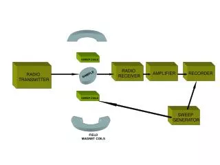
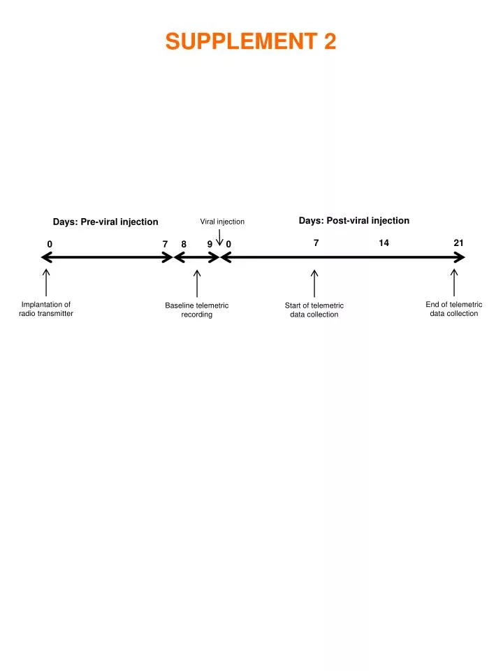
Days: Post-viral injection. Days: Pre-viral injection. Viral injection. 7. 14. 21. 0. 7. 8. 9. 0. Implantation of radio transmitter. Baseline telemetric recording. Start of telemetric d ata collection. End of telemetric d ata collection. SUPPLEMENT 2. 70. SHR. 60. WR. 50.

E N D
Days: Post-viral injection Days: Pre-viral injection Viral injection 7 14 21 0 7 8 9 0 Implantation of radio transmitter Baseline telemetric recording Start of telemetric data collection End of telemetric data collection SUPPLEMENT 2
70 SHR 60 WR 50 40 Mean Cell Count 30 20 10 0 DBH eGFP SUPPLEMENT 3 Numbers of viral vector transduced and dopamine-b-hydroxylase (DBH) immunopositive neurons were not different between Wistar (WR) and spontaneously hypertensive rats (SHR) To compare cell counts of eGFP expressing neurons (i.e. efficacy of transduction) and norepinephrine-containing (DBH-positive) neurons between WR and SHR, immunolabeled neurons were counted in every fifth section in each WR and SHR animal. No difference was found in the number of eGFP expressing neurons between WR and SHR (P=0.44; ANOVA, based on counts from 6 animals) suggesting that the efficacy of transduction was similar between rat strains. Moreover, we found no difference in the numbers of DBH-positive neurons in SHR and WR (P= 0.5; ANOVA, based on counts from 6 animals). Difference in numbers of DBH- and eGFP-expressing neurons may reflect the limitations of immunocytochemical sensitivity and possible inclusion of dorsal vagal motoneurones at the NTS-dorsal vagal motor nucleus boundary.
Wistar SHR SUPPLEMENT 4 Membrane potential (mV) Resting membrane potential in A2 neurons in Wistar and spontaneously hypertensive rats (SHR) In order to ascertain the membrane potential and input resistance of A2 neurones in SHR and Wistar rats, we prepared organotypic brainstem slice cultures of the medulla oblongata at the level of the caudal NTS and expressed EGFP in A2 neurones as described previously1. EGFP-fluorescent A2 neurones were then patched and recorded in whole cell mode using established protocols2. There was no statistical difference in the resting membrane potentials between these rat strains: Wistar: -57.4 + 3 mV; SHR: -59.4 + 4 mV (P=0.71, Student’s t-test; n=5 each). Likewise, membrane input resistance was not different between the two rat strains (P>0.3). It may be concluded that differences in basic membrane properties of the A2 neurons between SHR and WR cannot account for different physiological outcomes of our in vivo experiments. References 1. Teschemacher, AG, Wang S, Lonergan T, Duale H, Waki H, Paton, JFR & Kasparov S. Targeting specific neuronal populations using adeno- and lentiviral vectors: applications for imaging and studies of cell function. Exp Physiol. 90:61-69, 2005 2. Wang S, Teschemacher, AG, Paton JFR, & Kasparov S. Mechanism of nitric oxide action on inhibitory GABAergic signaling within the nucleus tractus solitarii. FASEB Journal 20 online (9):1537-1539, 2006.