Download
1 / 41
540 likes | 1.68k Views
INVASIVE PROCEDURES. by DR J JEEBODH . AIM OF PRESENTATION. Briefly discuss options available for prenatal diagnosis Briefly discuss therapeutic options Procedure/technique Complications Limitations. DIAGNOSTIC. Amniocentesis Chorionic villus sampling

E N D
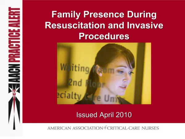
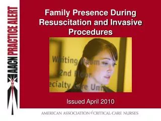
INVASIVE PROCEDURES by DR J JEEBODH
AIM OF PRESENTATION • Briefly discuss options available for prenatal diagnosis • Briefly discuss therapeutic options • Procedure/technique • Complications • Limitations
DIAGNOSTIC • Amniocentesis • Chorionic villus sampling • Cordocentesis and other methods of FBS • Fetal tissue biopsies • Coelocentesis • Fetoscopy
AMNIOCENTESIS Removal of fluid containing fetal cells and biochemical products from amniotic cavity Cells in amniotic fluid are desquamated from fetal skin & sloughed from GIT,urogenital & resp tract and amnion
INDICATIONS • CHR and DNA analysis • Biochemistry • Fetal infection • Lung maturity • Chorioamnionitis • Obstetric cholestasis • Therapy
PROCEDURE • Performed more than 15weeks & up to 18 weeks • Consent and counselling • Sonar • Transabdominal approach • Asepsis • Direct and continuous ultrasound guidance • 22 gauge spinal needle • Avoid fetus , placenta and cord
EARLY AMNIOCENTESIS • Less than 15 weeks • Only 10 to 12 ml • Success between 12 to 14 weeks ?95% • Fewer cells to culture • Advantages • Complications : More than post midtrimester amnio, failure rate, fetal loss
COMPLICATIONS • Fetal loss • Pregnancy complications – abdo pain, amniotic fluid leakage , vaginal bleeding • Chronic leakage • Orthopaedic abnormalities • Fetal trauma • Rhesus alloimmunisation
PREDISPOSING FACTORS TO SPONTANEOUS ABORTIONS POST AMNIOCENTESIS • History of previous spontaneous or induced abortion • Bleeding in current pregnancy • Age > 40 yrs • 3 or > 1st trimester abortions • 2nd trimester miscarriage or TOP • Obstetrician/operator experience
AMNIOCENTESIS & TWINS • Single needle technique better than double • Needle into proximal sac – aspirate • Advanced into 2nd sac through membrane under direct vision • Concerns • DO NOT USE DYES • Fetal loss risk
CVS AND PLACENTAL BIOPSY • Chorion frondosum or placental tissue sampled • Alternative to amniotic fluid and FBS for prenatal diagnosis of genetic disorders • Villi excellent source of DNA • Tissue obtained consists of syncytiotrophoblast (outer non-dividing cells) and cytotrophoblast (rapidly dividing cells) Cytogenetic results in 48hours & final culture in 7d
INDICATIONS FOR CVS • Maternal age at 35 yrs or more at conception • Previous child with aneuploidy • Parent who is a carrier of a balanced translocation or other CHR abnormalities • Autosomal recessive disease • Women who are carriers of sex – linked disease
PROCEDURE • Between 11 to 14 weeks • Transabdominal or transcervical (less frequently) • Needle into placenta under continuous simultaneous ultrasound guidance • Placental biopsies done in 2nd and 3rd trimesters
COMPLICATIONS • PROCEDURE RELATED Miscarriage Limb reduction Risk of late termination • DIAGNOSIS RELATED More cytogenetic ambiguous results Mosaicism & other variants(1%) – false negative results
CVS AND LIMB DEFICIENCES • Risk and severity of limb deficiency appear to be associated with timing of CVS • Overall risk for transverse limb deficiences 0.03% - 0.1% (1 in 3000 to 1 in 1000) • Less than 10 weeks : 0.2% & more proximal limb deficiences and orofacial defects • At or greater than 10 weeks : 0.07% & most limited to digits • Possible mechanism some form of vascular disruption
CVS AND TWINS • Loss rate similar to amnio • Concerns • Techniques to reduce contamination • Advantages • Disadvantages
FETAL BLOOD SAMPLING DIAGNOSTIC CHR abnormalities DNA abnormalities / single gene defects Fetal anaemia Fetal thrombocytopaenia Fetal hypoxia / acidosis Fetal infection THERAPEUTIC Anaemia , thrombocytopaenia , drug administration , fetacide
PROCEDURE OPTIONS SAMLING SITE 1. Fetal heart 2. Fetal intrahepatic vessels 3. Umbilical cord (cordocentesis) -Easiest site to puncture is 1cm from cord insertion into placenta (avoid free loop) -More than 20 weeks : 5ml -Less than 20 weeks : caution with volume removed
COMPLICATIONS • FETAL - bleeding or haematoma • fetal bradycardia • Chorioamnionitis • Placental abruption • Amniotic fluid leakage or ROM • Death • Disability in survivors • Transmission of maternal infection
MATERNAL • Alloimmunisation • Chorioamnionitis • Maternal trauma • Possibilty of emergency delivery Post procedure pregnancy loss rate :1% • Tranfusion carries higher procedure related risk than blood sampling • Operator skill important
COELOCENTESIS • Earlier than CVS • Coelomic space between amniotic membrane and uterine cavity • Under re-evaluation • 1 to 2.5ml of amniotic fluid (vol by 9 weeks ±5 to 6ml) • Cells mostly haemopoetic in origin • 90% of cells viable before 7 weeks • 95% success at 7 weeks
FETAL TISSUE BIOPSIES • Need for diagnosis of disorders not amenable to molecular approaches • Indications are possible or potentially lethal or severely handicapping conditions in fetus affecting skin, liver, muscle & occasionally for diagnosis of fetal tumours • Options : fetoscopic directed biopsies ,ultrasound guided aspiration or ‘tru-cut’ technique
FETAL SKIN BIOPSIES • Only a few major dermatological disorders associated with CHR abnormalities or enzyme defects detectable on either amniotic fluid or chorionic villi • Ultrasound visualisation useless in majority of serious cutaneous abnormalities • Actual visualisation of skin and histology only way to make diagnosis • Obtained under direct visualisation using fetoscopy or ultrasound guidance • Heal with no scar formation • Complications : ROM ,bleeding , infection , miscarriage
FETAL LIVER BIOPSIES • Successfully used for prenatal diagnosis of : - ornithine transcarbamylase deficiency - carbamyl phosphate synthetase deficiency - Von Gierke’s disease - Primary hyperoxaluria type 1 • Needle or coring biopsy instrument inserted into RUQ of fetal abdomen
FETAL MUSCLE BIOPSIES • Majority of DMD currently diagnosed by molecular analysis of the gene using CVS • Either through detection of deletion mutation or by linkage analysis • Is possible that deletion mutation not found • Muscle biopsy allows finding of dystrophin • Complications : nerve damage & bleeding • ONLY USE IF INDICATED • EXPERIENCED OPERATOR
Other organs : lung , kidney • Tumours • Most indications DO NOT outweigh risk • Risk of pregnancy wastage relatively high • Biopsy when yield exceeds risk • Complex analysis of specimens
THERAPEUTIC INVASIVE PROCEDURES • Rhesus isoimmunisation • Platelet disorders • Drugs • Multiple pregnancy reduction and selective termination • Fetal shunts • Relief of polyhydramnios • Therapeutic amnioinfusion • Open fetal surgery • TTTS • Fetoscopy
MULTIFETAL PREGNANCY REDUCTION AND SELECTIVE TERMINATION • Obstetric outcome of triplets & higher numbers of fetuses significantly compromised • First trimester procedures for fetal number: MFPR • 2ND Trimester procedures for fetal abnormalities : selective termination
MFPR • Transvaginal aspiration or transabdominal KCL • Embryo selection - exclude abnormalities or aneuploidy - choice based on ease of approach - avoid embryo closest to cervix • Timing – 9 to 12 weeks
SELECTIVE TERMINATION OF ABNORMAL TWIN • Case selection critical • If placenta shared ,demise of one twin potentially places surviving co–twin at increased risk • KCL only in dichorionic twins • Umbilical cord ligation or occlusion in monochrionic twins • Risk of fetal loss
TTTS MANAGEMENT • Expectant • Medical no benefit • Serial amniodrainage • Septostomy with /without amniodrainge • Selective fetocide – ligation of umbilical cord/ cord coagulation, cord embolisation • Laser occlusion – fetoscopic directed laser ablation of vascular anastomoses
FETOSCOPIC LASER ABLATION • Cause-orientated approach • Fetal complications • Maternal complications • Fetal survival • Risk of neurological sequelae
FETAL SHUNTS • Can perform antenatal drainage of pathological collections of fluids in fetus • Direct needling or shunting • In theory, drainage of abnormal fluid collections possible from fetal abdo,brain,chest,renal tract • Pleural effusions – pleuroamniotic shunt • Vesicoamniotic shunts in obstructive uropathy or to decompress a dilated urinary tract
BLOOD TRANSFUSIONS • Intraperitoneal(less preferred) or intravascular • INTRAPERITONEAL - blood absorbed via lymph vessels - 10% of cells absorbed each day by non-hydropic fetus - absorption in hydropic fetus poor - disadvantages : slow correction of HB,increased risk of trauma, can obstruct cardiac return, lower survival rates than intravascular
BLOOD TRANSFUSIONS CONTINUED • INTRAVASCULAR - Umbilical vein - 22 gauge needle - at insertion or free loop volume determined by Hct and gest age • When to transfuse? Hct < 30 % ( <25th percentile for GA) OR • Vol = (weeks in gestation – 20) x 10ml
THERAPEUTIC AMNIOCENTESIS • Transabdominal • Decrease amniotic fluid under sonar guidance • Prolong gestation & improve survival • Aim : DVP <8cm or AFI normal range • Complication rate 1.5% - PROM, chorioamnionitis, abruptio, membranous detachment • Maximum 3litres • TNT patch
AMNIOINFUSION • INDICATION : Improve visualisation of fetal anatomy or severe oligohydramnios / anhydramnios • FLUIDS: physiological solution- N/S, RL,5% gluc • VOL: Minimum needed to improve visualisation • COMPLICATIONS
CONCLUSION • Risk of HIV transmission • Counseling – Procedure related risks, complications, limitations • Individually tailored risk assessment before procedure