Download

1 / 30
320 likes | 884 Views
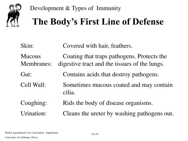
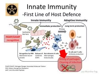
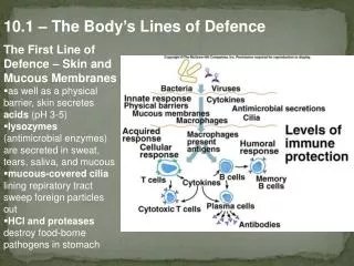
The First Line of Defence. The first line of defence consists of physical and chemical barriers to prevent the entry: Skin Mucus Cillia Chemical barriers and Other bodily secretions. 4.2 identify antigens as molecules that trigger the immune response. Anti = Antibody

E N D
The first line of defence consists of physical and chemical barriers to prevent the entry: • Skin • Mucus • Cillia • Chemical barriers and • Other bodily secretions
4.2 identify antigens as molecules that trigger the immune response Anti = Antibody -gen = generator
Antigens are often part of the outer coating of a bacterium or virus. The antigen is recognised by the body as not being part of itself, and antibodies are released to attach to the antigen. Antigens on a bacterium
Inflammation response If a pathogen (eg bacteria) is successful in penetrating the first line barriers, injured cells let off a chemical alarm such as histamines. Inflamation- This causes blood vessels to dilate, increasing blood flow to the area, and increasing the permeability of the vessels. The area becomes swollen, red and painful.
Lymphatic System • The lymphatic system returns intercellular fluid to the blood system, filters cell debris and produces white blood cells responsible for the immune response. • Includes: Spleen, thymus, tonsils and lymph nodes and vessels.
Phagocytosis • Some white blood cells, called macrophages and neutrophils, can very easily change their shape so that they flow around particles and completely enclose them within their cell, where they are broken up by cell enzymes. This is called phagocytosis.
Animation - Phagocytosis • http://www.youtube.com/watch?v=VAhM9OxZDkU
Cell death to seal off pathogens • For some pathogens, macrophages and lymphocytes completely surround a pathogen so that it is enclosed in a cyst. The white cells involved die so that the pathogen is isolated from its food supply and also dies.
3rd Line of Defence • identify the components of the immune response: —antibodies —T cells —B cells
Antibodies are proteins, called immunoglobulins, which are produced in response to the presence of an antigen in the body. When the appropriate B cells are activated they form plasma cells that produce antibodies, the antigen binding sites of which match the shape of the antigen they are specific for. These antibodies then seek out the antigen and bind to a part of it, forming the antigen–antibody complex, which causes the deactivation of the antigen. There are a number of ways in which the antigen can be destroyed, including - immobilising it, blocking and neutralising the active binding site of the antigen, or by causing the antigen– antibody complex to clump together, making them easier to eliminate by phagocytosis. Antibodies
B cell T cell Cancer cell
T cell - Another type of lymphocyte. - Differentiate in thymus gland. - Thus ‘T cell’. - Remain inactive in blood & lymph until they detect an antigen. • Antigen binds to T cell, activating cloning mechanism. - T cells control cell-mediated response. - Various types of T cells destroy antigen / foreign cell.
Cytotoxic T cells: - Carry antigens. - Remove foreign proteins from body. - Anything recognised as ‘non self’. - Bacteria. - Transplants. Natural killer cells: - Special cytotoxic T cells. - Destroy abnormal host cells. Eg. Cancer cells, viruses. Helper T cells: - Secrete interleukins. - Regulate cytotoxic T cell & B cell functions. - Inducer T cells, suppressor T cells: - Regulate T & B cells. - Start & stop production and action. Memory T cells: - Recognise antigen when reappearing. - Have helper T cell function. - Quick supply of antibody to antigen.
Immune Response - Antigen enters body. - Travels via blood to lymphoid tissue (lymph node or spleen). - Ingested, processed by macrophage. - Displays fragments of antigen on outer membrane. - Recognised by helper T cells & B cells. - Interact through cytokines (secreted by T cells & macrophages) - Signal other cells to initiate immune response. Eg. B cell to transform into plasma cell. Interaction between B & T lymphocytes: - Attack same antigen. - ‘Helper T cells’ stimulate B & T cells to clone.
Mechanisms 2 proposed mechanisms for interaction of B & T cells. Mechanism 1: - T cell produces soluble factor after interaction with antigen. - B cell reacts with factor & specific antigen. - B cell becomes functional antibody producing cell. Mechanism 2: - Based on cell contact between T & B cell. - Contact arises from interaction with antigen. - Contact allows T cell to signal B cell to become functional. - Antibody producing cell.
Collaboration of Cells - Close to each other. - Regulated by cytokines. - Proteins/polysaccharides. - Secreted by T cells & macrophages. - Signal other cells to initiate immune response. Mechanisms allowing interaction of B & T lymphocytes: - T lymphocytes help B lymphocytes. - Helper T cells (Th cells). - If B cell has antigen on surface, risk that T cell will recognise antigen & attack it & B cell. - T cell able to recognise ‘self’ molecules.
1 2