Download
1 / 32
320 likes | 321 Views
This chapter explores the classical traits used to identify and group prokaryotic microorganisms, such as cell wall thickness, presence of endospores, cell morphology, and cell arrangements. It also discusses the genetic traits used for classification, including conserved gene sequences like 16S rRNA. The similarities and differences between Archaea and Bacteria are examined, highlighting their cell walls, ribosomal RNA sequences, and other distinctive traits. Extremophiles, such as thermophiles and halophiles, are also discussed.

E N D
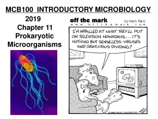
MCB100 INTRODUCTORY MICROBIOLOGY 2019 Chapter 11 Prokaryotic Microorganisms
Chapter 11 Prokaryotic Organisms Classical Traits Used to Identify and Group Microorganisms - Presence of a thick vs. thin cell wall – the Gram Stain - Presence of endospores - Cell morphology (shape) - Cell Arrangements (group structure) - Biochemical Tests (metabolic pathways, enzymes) vs.
Classification Based On Microscopic Morphology (an old approach) Common cell shapes: Coccus: round berry shape Bacillus: rod shaped Vibrio: bent rod Spirillum: thick helical shape Spirochete: very skinny helical shape Coccobacillus: well gee.. it’s hard to say Common Cell Arrangements: Strepto: chains Staphylo: a bunch Tetrads: groups of 4 Sarcina: groups of 8 Diplo: two Palisades: V-shaped
Genetic traits used to classify microorganisms • Conserved gene sequences such as the 16S rRNA • All cellular organisms have ribosomes. • 16S rRNAs are about 1542 bases in length. • You can think of each base in the sequence as a trait.
Genetic traits used to classify microorganisms Conserved gene sequences – such as 16S rRNA Above: How gene sequence comparisons work. The more closely related two organisms are, the fewer changes will be seen in the sequences. Right: Neighbor-joining tree for the 16S rRNA sequences for Lactobacillus species. From: scielo.cl
Classification Based on Gene Sequences (especially rRNA genes) Analysis of rRNA sequence data is useful for determining relationships between organisms having ribosomes (everything except viruses, viroids and prions). Molecular Classification (based on gene sequences, especially rRNA genes) - Based on genotypic data rather than phenotypes - Data is analyzed by a computer using mathematical algorithms that remove human bias. - Pioneering work was done by Carl Woese atIllinois
The Archaea and Bacteria – Similarities - Prokaryotic cell – no nuclear membrane, one circular chromosome - 70S ribosomes - most have cell walls - some species stain purple and others end up red in the Gram stain Methanopyrus Differences Between the Archaea and The Bacteria - Ribosomal RNA sequence data - Archaeal cell walls lack peptidoglycan (no N-acetyl muramic acid). - Archaeal cytoplasmic membrane lipids have branched chain fatty acids that may extend through the membrane. - The initial amino acid in protein synthesis in Archaea is methionine rather than formyl-methionine, which is used in bacteria. - Archaeal RNA polymerase is similar to eukaryotic RNA pol II. - Archaeal flagella are simple protein threads that rotate like those of bacteria, but they are very different. Archaea flagella are not hollow tubes, they are solid. Assembly differs from bacterial flagella too.
Some Traits of the Archaea 1) Archaeal cell walls are chemically different from Bacterial cell walls. - resistant to Lysozyme, Penicillins and Vancomycin - vary among taxa(In the Gram stain, some are purple and others red.) - composed of a variety of compounds (proteins, glycoproteins, lipoproteins and polysaccharides but not peptidoglycan, they don’t use N-acetyl muramic acid) Pseudomurein Pseudomurein is a cell wall polymer found in some species of Archaea that is similar to peptidoglycan in that it consists of sugar chains that are cross linked by amino acid chains. Pseudomurein differs from peptidoglycan in that it contains N-acetyl-L-talosaminuronic acid instead of N-acetyl-muramic acid, the cross-linking chains contain only L-amino acids, and the linkages of the sugar polymer are B(13) rather than the B(14) linkages that are seen in bacterial peptidoglycan. 2) There are currently no Archaea known to be human or animal pathogens. 3) Archaea include Methanogens and Extremophiles
Bacterial Peptidoglycan (Murein) vs. Archaeal Pseudomurein Bacterial Peptidoglycancontains n-acetyl muramic acidsugars linked by beta - 1,4 bondspeptide includes unusual amino acidssuch as D – alanine, D – glutamic acid,diaminopimelic acid and ornithine Archael Cell Wallscontain n-acetyl talosaminuronic acidsugars linked by beta - 1,3 bondspeptide includes only ordinaryL – amino acids
Extremophiles – Thermophiles Thermophiles grow at temperatures over 45oC. Hyperthermophiles grow at temperatures over 80oC. Examples of thermophilic Archaea Sulfolobus grows best at 75oC at a pH of 2.5, and is an obligate aerobe. Pyrodictium grows best at 105oC at a pH of 6, and is anaerobic (note: not all thermophiles are Archaea, not all Archaea are thermophiles) Hot Springs
Extremophiles – Halophiles - Halophiles grow in extremely salty environments - Their habitats are more than 9% NaCl (sea water is 0.9%) Some survive in 35% NaCl Many make red-orange pigments that absorb light energy and protect from UV. Example: Halobacterium, Halococcus - Use sunlight to produce a PMF that is used to make ATP - Lack chlorophylls or bacteriochlorophylls Makes Bacteriorhodopsins (purple-red pigments, rhodo = red) - Rhodopsinprotein helps protect Halococcus from the extreme salinity of their natural environments Salt Evaporation Ponds near San Francisco. The bright red colors are caused by pigments in Halococcus.
Methanogens Methanogens are obligate anaerobes that produce methane - CH4 . Substrates for methanogenesis include: CO2 + H2, methanol and various organic acids Some methanogens are thermophiles - Methanogens convert organic wastes in pond, lake and ocean sediments into methane and CO2. - Methanogens make gas in the gut of cows and grass eating animals. - Methanogens play an important role in the anaerobic digestion of organic wastes in sewage treatment. Examples: Methanopyrus (Pyro = fire) Methanobacterium
Bacteria Currently grouped into about 30 phyla based on 16S rRNA sequence data. Deeply Branching Bacteria (thought to be similar to very earliest life forms) Examples: Aquificaeles species Hyperthermophyllic chemoautotrophic anaerobes Derive energy and carbon from inorganic sources (CO2 + H2) Habitat includes hot mineral springs Deinococcus radiodurans Multiple copies of the chromosome and radiation absorbing pigments allow it to endure exposure to extremely high doses of ionizing radiation. (They have an outer membrane like Gram-negative bacteria but stain purple in the Gram stain.) Filaments of chemolithotrophic thermophilic Aquificales bacteria at Mammoth Terraces in Yellowstone (With butterfly)Photo from A.L. Reysenbach, Portland State University
Phototrophic Bacteria - Blue-green bacteria (Cyanobacteria, blue-green algae) - Green sulfur bacteria - Green non-sulfur bacteria - Purple sulfur bacteria - Purple non-sulfur bacteria Cyanobacteria Chlorobium carries out anoxygenic photosynthesis under anaerobic conditions.
Oxygenic Photosynthesis in Cyanobacteria 12H2O + 6CO2 C6H12O6 + 6H2O + 6O2 (Plant chloroplasts are thought to have evolved from endosymbiotic cyanobacteria.) Cyanobacteria Beggiatoa Anoxygenic Photosynthesis in Sulfur Bacteria 12H2S + 6CO2 C6H12O6 + 6H2O + 12S0
GRAM POSITIVE BACTERIA (Gram-positive bacteria have a thick cell wall composed of peptidoglycan with teichoic acids and no outer membrane or periplasmic space.) Low G + C Gram-positive Bacteria(Firmicutes) (This means that the DNA contains more A=T basepairs than G=C basepairs.) Clostridium Bacillus Mycoplasma Listeria Lactobacillus Streptococcus Enterococcus Staphylococcus High G + C Gram-positive Bacteria(Actinobacteria) (This means that the DNA contains more G=C basepairs than A=T basepairs.) Corynebacterium Mycobacterium Actinomycetes
Clostridium • (Clostridium means club-shaped.) • - Anaerobes (many are obligate anaerobes that die • in the presence of O2) • - Produce heat resistant endospores • - They have a wide variety of fermentative pathways • such as acetone-butanol production • Some species produce and excrete potent toxins • About 1/3 of the bacteria in the human large intestine are Clostridium species • Examples • Clostridium botulinum • Clostridium tetani • Clostridium perfringens • Clostridium difficile
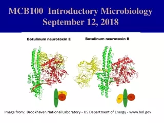
Clostridium botulinum • Anaerobic, spore forming, gram + rod • Botulism: severe form of food poisoning • Improperly canned non-acidic food • Neurotoxin • Headache, double vision, flaccid paralysis
Clostridium botulinum - natural habitat is anaerobic sediments in ponds and soil - produces a potent neurotoxin that causes flaccid paralysis - has been involved in large duck die offs - grows in contaminated canned food - most cases of botulism are due to ingestion of toxin - botulism toxin is denatured by cooking - may infect the stomachs of infants and produce toxin in vivo - spores are common in honey - botox is used therapeutically to relax facial muscles - A-B type toxin enters host cells and enzymatically inactivates neurotransmitter releasing mechanisms Clostridium tetani - natural habitat is anaerobic soil layers and sediments - produces a toxin that causes rigid paralysis - infects wounds and produces toxin in vivo - tetanus vaccine is a toxoid that provides immunity to toxin
Tetanus • Clostridium tetani • Gram positive rod, spore former, anaerobic • Predilection for deep, soil contaminated, wounds • Tetanospasmin • Prolonged contraction of muscles • Voluntary muscles do not relax
Tetanus • Incubation period: 3 days – 3 weeks • Lock Jaw: prolonged contraction of massiter • Vaccine is a toxoid (inactivated tetanospasmin) • Antitoxin available • Protect patients from stimuli • Untreated mortality rate of 40-80% • Medical procedures can cause
Factors Affecting the Development of Wound Infections Virulence of contaminating organism Size of contaminating dose Extent of tissue damage at wound site Health status of wounded person An infection that is limited to the skin, nails, mucous membranes, hair etc. is called a topical infection. An infection that spreads throughout the body is called a systemic infection.
Clostridium perfringens - spores are common in soil - wound infection - causes gas gangrene - produces a number of necrotizing toxins that kill tissues Clostridium difficile - may be found in low numbers in the human gut - naturally resistant to several antibiotics - may multiply in patients on antibiotic therapy - produces toxins - may cause antibiotic induced diarrhea
Clostridium perfringens Anaerobic, spore forming, Gram positive rod Found in soil, animal feces, meat & poultry contaminated at slaughter Causes gas gangrene if it infects a wound Also can cause food borne infection Exotoxin produced in intestines after ingesting contaminated food Symptoms include: abdominal pain, diarrhea
Clostridium difficile • C. difficile is a major cause of colitis following antibiotic use, especially in hospitals. • C. diff. makes two toxins cleverly called: A&B. • Tissue destruction in the colon causes formation of a plaque-like pseudomembrane. • Symptoms are: watery diarrhea, with no blood. • To treat, stop antibiotic therapy, give probiotics. Early stages of C. difficile infection images from trialx.com
Clostridium difficile colonizes the colon but generally does not become very numerous because it does not compete well with the natural gut flora. C. difficile is naturally resistant to several antibiotics. Antibiotic therapy allows the bacteria to multiply to high numbers. C. difficile toxins cause diarrheal disease.
Ruminococcus flavefaciens Ruminococcus flavefaciens is a Gram +, anaerobic, gut microorganism now grouped in the Clostridia class. It helps ruminantanimals digest cellulose. cellulose