Download
1 / 39
390 likes | 620 Views
Ophthalmoscopy. David Kinshuck. This session 1. Over the next hour Ophthalmoscopy in a younger person Need to practice for older patients 15 minute introduction Practice on each other for 40 minutes 3 minute summary If time we may cover refraction. This session 2. Very briefly

E N D
Ophthalmoscopy David Kinshuck
This session 1 • Over the next hour • Ophthalmoscopy in a younger person • Need to practice for older patients • 15 minute introduction • Practice on each other for 40 minutes • 3 minute summary • If time we may cover refraction
This session 2 • Very briefly • External examination • Test pupils • Test visual fields • In more detail, ophthalmoscopy itself • Red reflex • Examine disc • Rest of retina
Ophthalmoscope controls 1 • Play with ophthalmoscope • Work out what the controls are for
Pupils • Pupils(needs Flash player installed)
Ref reflex technique • Check red reflex from 10 cm, focusing on iris • +3.00 lens? • Actually focus on the pupil margin • Brightest light in ophthalmoscope • Stay on same side of patient for both eyes • Room lights dim
www.occhioallaretina.it/Immagini/leucocoria.JPG http://oftalmologo.bravepages.com/Leucocoria%201.jpg ‘white’ reflex ?retinoblastoma
Ophthalmoscopy 2 • What is your refraction • What is the patient’s refraction • ……what lens do you use in the ophth.? • Light travels from patient retina to your retina
Ophthalmoscopy 3 • Distance: closer you are, the greater the visual field..1” away is best • Pupil size for ophthalmoscope beam must match patient’s pupil size (otherwise get too much reflection) • Too bright…pupil goes small • Too dim..not see • Need room lights off/dim
Ophthalmoscopy 4 the closer you get, the larger area of retina seen at one time; Look in from 2 cm away
Ophthalmoscopy 5 Match beam diameter with pupil size.. not too wide Just right, no reflection Too wide a beam leads to increased reflection
Ophthalmoscopy 6 Match beam diameter with pupil size..not too narrow Too narrow a beam…too little retina is seen
Ophthalmoscopy 7 If beam is too bright, pupil goes small (then get a reflection). Too dim, won’t see anything!
Ophthalmoscopy 8 Patient looks up, doctor down (more comfortable/quicker)
Ophthalmoscopy 9 Index finger on focus dial, Thumb on brightness control (rheostat)
Ophthalmoscopy 10 Consider using middle finger to gently touch cheek …steadies ophthalmoscope and prevents banging into eye
Ophthalmoscopy 11 Consider resting ophthalmoscope on thumb (which rests over eyebrow) to steady ophthalmoscope and prevent banging into the eye
Ophthalmoscopy 12 Hold ophthalmoscope immediately adjacent to your own (observer’s) eye to get the best view.
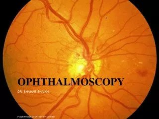
Ophthalmoscopy 13 Examine optic first (looking medially 15o. Otherwise, as soon as light hits the fovea, pupil will go smaller, making examination more difficult.
Ophthalmoscopy 14 Use the grid to locate the fovea (the centre of the macula).
What is the most important part of the retina for the none-ophthalmologist? • The optic nerve …. • Papilloedema, raised intracrainial pressure & many other conditions
How to find the optic nerve Look medially 15o …optic nerve, pituitary, optic tract/cortex lie on the same 15o axis
How to find the optic disc Vessels point to the optic nerve, so find a vessel fork and move towards optic disc
summary • What did you find easy • Hard • Red reflex +3.0 d • Retina focus..your prescription + patient’s • Dim room light • Match beam diameter with pupil size at 2 cm • Get close…2 cm….larger area of retina visible • Beam not too bright otherwise pupil goes small • Patient looks up 15o • Rest finger on cheek? • Follow vessel branching towards the optic disc • Disc is 15o medial • Use grid in ophthalmoscope to locate fovea
Refraction 1 Which lens is needed for which eye?
Refraction 2 Use the appropriate lens for the eye Add to your own spectacle prescription for ophthalmoscopy
Refraction 3: how to find the plus lens Focus on something very close
Refraction 4: how to find patient’s spectacle prescription Hypermetropic lens magnifies Myopic lens makes everything appear smaller Emmetropic
Refraction 5: examining high myopes For myopes there is only one focal plane…any further back and you will be out of focus. This is unlike emmetropes: you will be in focus even if you examine from a distance (although only a tiny amount of retina will be visible). Also, unless you have an excellent ophthalmoscope, you may be best examining by looking through the patient’s own spectacles.