Download
1 / 13
130 likes | 261 Views
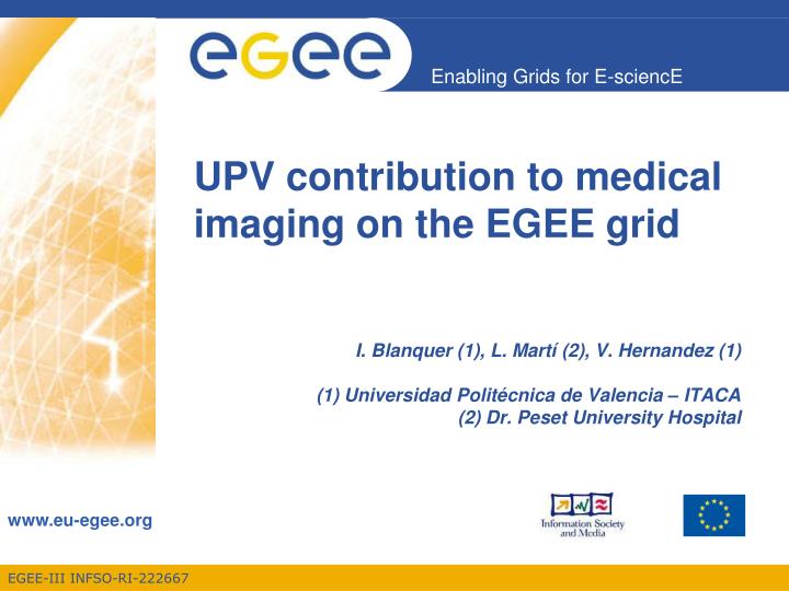
UPV contribution to medical imaging on the EGEE grid. I. Blanquer (1), L. Martí (2), V . Hernandez (1) (1) Universidad Politécnica de Valencia – ITACA (2) Dr. Peset University Hospital. Contents. Objective of UPV’s Activities in Medical Imaging for EGEE.

E N D
UPV contribution to medical imaging on the EGEE grid I. Blanquer (1), L. Martí (2), V. Hernandez (1) (1) Universidad Politécnica de Valencia – ITACA (2) Dr. Peset University Hospital
Contents Objective of UPV’s Activities in Medical Imaging for EGEE. The TRENCADIS Middleware and its Connection to EGEE. DICOM Structured Reporting. Work in Progress.
Objective of UPV’s Activities in Medical Imaging for EGEE • UPV’s Participates in the Biomed Cluster (TNA4.2.2) in the LS3 (Support for Selected Services) and LS4 (Life Science Application Porting Support) Activities. • The Focus of the UPV in Those Activities is on the Support of Structured Radiology Reports and Fostering the Use of AMGA • Use of Semantic Structuring of Radiology Reports will Also Increase the Exploitation of Knowledge. • A Key Issue is the Representation of the Metadata From Image Diagnosis Structured Reports. • It will Complement DICOM MDM and TRENCADIS, Focused on the Images.
TRENCADIS Middleware: Concept • Towards a Grid Environment for Processing and Sharing DICOM Objects • TRENCADIS Aims at the Development of a Middleware to Create Virtual Repositories of DICOM Images and Reports. • It Uses a Semantic Model for Organising the Data. • Data is Encrypted and Decrypted to Ensure Privacy Protection. • High-Performance Services are Included with the System. • Architecture Totally Based on WSRF. • Objective: Creation of Virtual Shared Repositories of Medical Images. • Complementary to PACS. • Intended Mainly for Research and Training. • Multicentric and Multiuser. • Data to be Shared is Explicitly Selected. • Data is Pseudoanonimised Before Entering in the System.
Semantic Organisation Users Organise Themselves on Virtual Communities. From the Studies Available, Only Those Matching the Selection Criteria of the Virtual Community Profile are Accessible. From the Images Available to a Virtual Community, a User Can Create an Experiment with the Studies Matching a Set of Restrictions. From this Experiment, More Detailed Views can be Obtained. The Criteria for the Selection of the Relevant Information Relies on the DICOM Tags of the Image and the Structured Report. TRENCADIS Middleware: Data Model View: (e.g. Patients Between 1 and 2 Years with hetrogeneous Supratentorial Findings) Experiment: (e.g Neuroblastoma) User Comunity (e.g. Paediatric Neuroimaging) Global Database: All the Images and Reports Shared
TRENCADIS Middleware: Data Model • Seven Templates Have Been Generated By the Experts • Report for the Staging of Malignant Liver Neoplasia, Small and Non-Small Cell Lung Cancer and Intraaxial Tumours of Central Nervous System. • Follow-up Reports for Liver Metastasis, Lung Carcinoma and Intraaxial Tumours of Central Nervous System. • The Reports are Structured and Coded Using the Rules of DICOM-SR. • Standard Coding (Mainly DICOM) Has Been Used When Possible, Following the DICOM-SR Rules To Introduce New Coding Schemas. • The Reports Generate Automatically the TNM Staging Code (From the Radiological Information) in the Cases of Liver and Lung.
TRENCADIS Middleware: Connection to EGEE • TRENCADIS Uses DifferentServicesCommontogLite and EGEE • VOMS AuthorisationMechanism. • gLite HTC and HPC GatewaysfortheMassiveSubmission of Jobs. • TRENCADIS Uses itsOwnIndexing, IS and Storage Systems
Conventional Report vs. Structured Report Observation of an Irregular EspiculatedMass of 7mm. of Maximum Diameter, Located on the inter-quadrant inferior line of the left breast. The Lesion Comprises Heterogeneous, Linear, grained and branched Microcalcifications Apparently a Malignant Lesion • Maglinancy • Based on: • Mass • Size: 7mm. • Shape: Irregular • Margins: Espiculated • Associated • Calcification • Tipus: Heterogeneous • Tipus: Branched • Distribution: Grouped. Free Text. There is no Structure Defined and Agreed and not Associated to Images. Structure and Lexicon Defined And Agreed. Associated to Images.
StructuredReport • Digital Report: • Properly Stored and Coded. • Fields Agreed by the Community. • Standard Lexicon and Semnatics. • Links to Images, Audio, Measures and Postprocessing Results. • Universal Medical Usage. • Based on Evidence. • Able to Reflect Experimental Results.
DICOM Structured Reporting: Concept • DICOM Structured Reporting Defines a DICOM-Compatible Format to Specify Structured Electronic Reports. • DICOM-SR Defines, Through DICOM Information Object Definition (IOD) Data Sets and Information Entity (IE) Data Element Objects, the Way to Specify Concepts and Coding Schemas. • DICOM-SR Defines Three IODs • Basic Text, Enhanced SR, Comprehensive SR. • IEs are Structured in a Tree-Shape • Each IE a Name / Value Pair, in Which the Name Comprises Three Items: Code Value, Code Schema and Code Meaning.
DICOM Structured Reporting: Templates • DICOM-SR Does not Define Which Fields Should be Included. • Templates are Being Provided and Agreed at DICOM • A Template is a Tree Structure with a Fixed set of IEs. • Defined in the “DICOM Content Mapping Resource" (DCMR) Supplement. • DICOM Working Group 15 Provides the Computer-Aided detection Templates for mammography (sup. 50), the chestCAD (sup. 65), patient history (sup.75), and breast report (sup. 79). • Other Templates for catheterization, ventriculography, intravascular US, US obstetrics and gynecology, vascular US, echocardiography, and fetal and pediatric echocardiography.
Work in Progress: Support for AMGA Databases DICOM SR Tree AMGA Schema Has Properties Has Properties • A DICOM Study is then a Pair DICOM-SR + DICOM Images. • DICOM Images Could be Huge and Comprise from one to Thousands of Files. • SEs are Good Storage Means (Either TRENCADIS DICOM Interaction Components or MDM SRM DICOM). • DICOM-SR Require a Relational or Documental Database. • AMGA is a Good Candidate to Standardise the Interface.
Work in Progress: Support for AMGA Databases • Develop three Tools • Translate a DICOM-SR Template into an AMGA Schema. • Insert a DICOM-SR File in an AMGA Database. • Retrieve a DICOM-SR File From an AMGA Database. • Benefit from the AMGA Features • Security and Authorisation. • Replication and Reliability. • Efficiency and Performance. • Standardisation and Availability of Services.