Download
1 / 20
210 likes | 256 Views
The Genetic Material. Brooker Chapters 2 & 9. Sept 6, 2007 BIO 184 Dr. Tom Peavy. What distinguishes living organisms from inanimate matter?. What are the requirements of “Genetic Material”?. Evidence that Genes Reside within Chromosomes. 1667- Anton van Leeuwenhoek (microscopy)

E N D
The Genetic Material Brooker Chapters 2 & 9 Sept 6, 2007 BIO 184 Dr. Tom Peavy
What distinguishes living organisms from inanimate matter?
What are the requirements of “Genetic Material”?
Evidence that Genes Reside within Chromosomes • 1667- Anton van Leeuwenhoek (microscopy) • Hypothesis: spermatozoa (“sperm animals”) enter the egg to achieve fertilization • Homunculus (spermists vs ovists)
Late 1800’s – microscopy studies • egg and sperm nuclei unite and contribute equally (e.g. frogs, sea urchins) • dyes used to stain the nucleus and observed long, threadlike bodies = Chromosomes (“colored bodies) • Mitosis described (nucleus is equally partitioned into daughter cells) • Sex Determination (♂ and ♀ chromosomes)
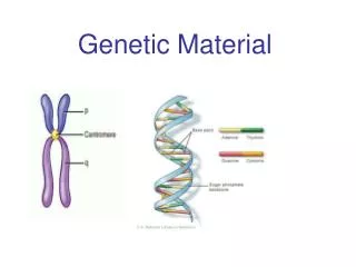
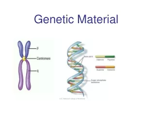
Homologous Chromosomes: The pair of chromosomes in a diploid individual that have the same overall genetic content. • One member of each homologous pair of chromosomes is inherited from each parent.
Chromosome theory of Inheritance (Sutton and Boveri 1902) • Chromosomes are in pairs and genes, or their alleles, are located on chromosomes • Homologous chromosomes separate during meiosis so that alleles are segregated • Meiotic products have one of each homologous chromosome but not both • Fertilization restores the pairs of chromosomes
Chromosomes • Approximately 40% DNA and 60% protein
Evidence for DNA as Genetic Material • Used simple experimental organisms to study question • Bacteria with single circular chromosome without a nucleus (prokaryotes) • Bacteriophage (“bacteria eaters”)
Frederick Griffith Experiments • In 1928, Griffith studied the bacterium Streptococcus pneumoniae • S. pneumoniae comes in two strains • S Smooth (strain IIIS) • Secretes a polysaccharide capsule (evades immune system) • Produce smooth colonies on solid media • R Rough (strain IIR) • Unable to secrete a capsule • Produce colonies with a rough appearance
The Experiments of Avery, MacLeod & McCarty • realized that Griffith’s observations could be used to identify the genetic material or “transforming principle” • Prepared cell extracts from type IIIS cells and added to type IIR cells for transformation in culture medium • Only the DNA enriched extract was able to convert type IIR into type IIIS • Further verification needed
Figure 9.3 Method • Allow sufficient time for the DNA to be taken up by the IIR • Add antibody that aggregates IIR bacteria (not transformed) • Gentle centrifugation • Plate remaining cells
Hershey and Chase Experiment (1952) • Studied the bacteriophage T2 • It is relatively simple since its composed of only two macromolecules • DNA and protein Inside the capsid Made up of protein Figure 9.4
Figure 9.5 Life cycle of the T2 bacteriophage
Figure 9.5 Life cycle of the T2 bacteriophage
Method • Used radioisotopes to distinguish DNA from proteins • 32P labels DNA specifically • 35S labels protein specifically • The two different Radioactively-labeled phages were used to infect non-radioactive Escherichia coli cells separately • After allowing sufficient time for infection to proceed, the residual phage particles were sheared off the cells • Phage ghosts and E. coli cells were separated • Radioactivity was monitored using a scintillation counter
Radioisotope Data (supernatant) Most of the 35S was found in the supernatant But only a small percentage of 32P