Download
1 / 98
1.1k likes | 1.28k Views
PORTAL HYPERTENSION. Rinsa Mary Oommen. 2002 Batch. Portal venous pressure above 10 mm of Hg or 30 cm of saline. Definition. Anatomy of portal circulation. 6.5cm long 0.8cm diameter Drains blood from Coeliac,SMA,IMA which supply to GIT spleen and pancreas. FORMATION.

E N D
PORTAL HYPERTENSION Rinsa Mary Oommen 2002 Batch
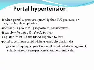
Portal venous pressure above 10 mm of Hg or 30 cm of saline Definition
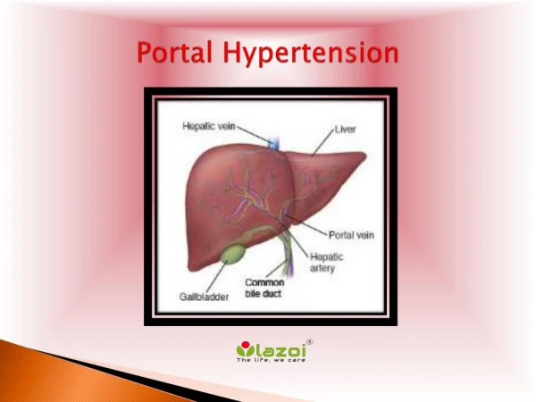
Anatomy of portal circulation • 6.5cm long • 0.8cm diameter • Drains blood from Coeliac,SMA,IMA which supply to GIT spleen and pancreas
FORMATION • Portal vein is formed by confluence of SMV and Splenic vein • behind the neck of pancreas • at the level of L1 or L2 Portal vein is formed by confluence of SMV and Splenic vein behind the neck of pancreas at the level of L1 or L2
Contd… • Liver is unique organ with dual blood supply 2/3rd-portal vein (nutrient rich blood) 1/3rd-hepatic artery (oxygenated blood from heart)
Contd… • In porta hepatis portal vein divides into two branches which goes to each lobe • Open into sinusoids • Empty into central veins • Sublobular veins • Interlobular veins • Hepatic vein -3 in no • IVC
Physiology of portal circulation • Hepatic blood flow – 1500 ml/min (25% of cardiac output) • Hepatic arterial auto regulatory or buffer response compensate in case of decreased hepatic portal bloodflow thus try to maintain hepatic blood flow in normal range
Contd… • Portal vein is avalvelessstructure resistance to PBF(at any level between right side of heart to splanchnic vessels) retrograde flow of blood from high pressure PVS to low pressure systemic venous circulation
Porto systemic collaterals-Sites Veins around 1)Cardiooesophageal jn (esophagogastricvarices) 2)Rectum(haemorrhoids) 3)Retroperitoneal veins 4)Periumbilical or abdominal wall collaterals
Clinical features 1)Hge from gastrooesophageal varices 2)Splenomegaly 3)Ascites 4)acute and chronic encephalopathy
While increased portal venous resistance initiates portal hypertension increased portal venous inflow secondary to hyperdynamic systemic circulation and splanchnic hyperemia maintains portal hypertension
Prehepatic • Portal or splenic vein thrombosis • Increased portal flow : Arteriovenous fistula, Massive splenomegaly from primary hematologic diseases
Hepatic Presinusoidal: • Schistosomiasis • Other periportal disorders (eg, primary biliary cirrhosis, sarcoidosis, congenital hepatic fibrosis), NCPF (Non-cirrhotic peri-portal fibrosis) Sinusoidal: • Cirrhosis (all etiologies) Post sinusoidal: • Veno-occlusive disease
Post hepatic • Hepatic vein thrombosis (Budd-Chiari syndrome) • Membranous obstruction of inferior venacava • Cardiac causes eg: constrictive pericarditis, restrictive cardiomyopathy.
Most common cause of portal hypertension in children – Portal vein thrombosis (PVT) • In PVT- Blood flow to liver maintained by collateral blood vessels ( cavernomatoustransformation of portal vein
LEFT SIDED PORTAL HYPERTENSION : • Etiology : Due to isolated splenic vein thrombosis (secondary to pancreatic inflammation or neoplasm) • CF- Gastric varices • Cure can be achieved by a splenectomy
EVALUATION • History: c/c alcoholism, hepatitis, complicated biliary disease • Examination : Signs of liver cell failure Splenomegaly
Investigations AIM To find cause of portal hypertension. To identify hepatic reserve. To define vascular anatomy. To find cause for GI hemorrhage
Laboratory tests • Hemogram : To diagnose hypersplenism • Liver function test : • Hypoalbuminemia indicates chronic liver disease. • Increased aspartate aminotransferase, alanine aminotransferase indicates ongoing hepatocellular necrosis. • Increased alkaline phosphatase indicates intra/extra hepatic cholestasis. • Increased PT and Bilirubin indicates hepatic decompensation. • Serology for hepatitis B and C virus.
Serum electrolytes : 1) Hypokalemia, 2)Hyponatremia and 3)Metabolic alkalosis secondary to hyperaldosteronism ,diarrhoea and recurrent emesis. consequences: Impairs tissue oxygen delivery Converts NH4cl to NH3- Hepatic encephalopathy
Contd… Liver biopsy : • For establishing etiology and assessing activity of liver disease. • Usually percutaneous route • Transjugular route if coagulopathy is present. Measurement of hepatic function reserve : Using child Pughclassification. Class determined on the basis of total points scored: 5 to 6 points - class A 7 to 9 points - class B 10 to 15 points - class C
HEPATIC VENOUS PRESSURE STUDY Portal pressure is indirectly estimated from hepatic venous wedge pressure Only use to differentiate between presinusoidal and sinusoidal or post sinusoidal causes of portal HT. • Portal vein anatomy : Esp before PSS operations • Selective visceral angiography • Now non invasive methods (Duplex USG, CT angiography,MR angiography) preferred.
CT Angiogram Portal venous anatomy
Diagnosis of bleeding • Nasogastric tube to confirm hematemesis • Upper GI endoscopy : Key procedure Idehtifying site of bleeding Classification of varices Assessment of gastric varices Estimation of risk of rebleed Treatment of bleeding varices
Cause of bleed in PHT • Oesophageal varices • Gastric varices • Portal hypertensive gastropathy
PHG • Only non variceal cause of portal hypertensive bleeding. • PHG mainly involves fundus and body of stomach. • Mild PHG - White reticular network with enclosed erythematous areas • Severe PHG – show granular mucosa and cherry red
VARICEAL HEMORRHAGE • Life threatening complication of portal hypertension • Mortality more in child-Pugh class C patients. • Esophageal varices develops in 60% of patients with cirrhosis and portal HT. • Only 30-50% of esophageal varices bleed • Predictors of variceal bleed : Child-Pugh class Variceal size Presence and severity of red wale markings (indicate epithelial thickness)
HISTORY: • Eck (1877) – Portocaval shunt • Crafoord & Frenckner(1939) – Endoscopic sclerotherapy • Sengstaken&blakemore(1950)- Balloon tamponade • Warren(1967) – Distal splenorenal shunt • Starzl(1967) – First successful liver transplantation • Inokuchi(1968) – Coronary caval shunt • Sugiura & Futagawa (1973) – Extensive esophagogastric devascularisation • Colapinto (1983) – TIPS in humans
MANAGEMENT • TREATMENT OF ACUTE VARICEAL BLEED • PREVENTION OF RECCURENT VARICEAL BLEEDING
Treatment of acute bleeding episode • INITIAL RESUSCITATION • PHARMACOTHERAPY • BALLOON TAMPONADE • ENDOSCOPIC TREATMENT • TRANSJUGULAR INTRAHEPATIC PORTOSYSTEMIC SHUNT (TIPS) • EMERGENCY SURGERY
RESUSCITATION • Establishment of an airway • Place largebore i.v lines – isotonic crystalloid solution or blood • Volume status assessed - monitor CVP and measure urine output • Full laboratory investigation done • FFP given if PT is prolonged(>3sec) • Monitor in ICU
Pharmacotherapy DRUGS USED Vasopressin Nitroglrcerine Somatostatin Octreotide Terlipresin Glypressin Metoclopramide pentagastrin
Vasopressin – most cmnly used S/E – Hypertension, PR, CO • Nitroglycerine – simultaneously infused • NEWER DRUGS : • Somatostatin 250mcg i.v bolus followed by 250mcg/hour • Octreotide (DOC) 50mcg i.v bolus followed by 25-50mcg/hour MOA – splanchnic blood flow by glucagon,VIP,substance p
Endoscopic treatment • Most commonly used therapy, controls bleeding in 85% of patients Methods - Sclerotherapy Band ligation
Sclerotherapy • Sclerosants used: Sodium tetradecyl sulphate, polidocanol, sodium morrhuate, ethanolamine oleate, absolute alcohol Cyanoacrylate glue for gastric varices • Technique : Intravariceal ligation Paravariceal ligation Combined
Complications : Fever,retrosternalchestpain, esophageal ulceration Rare – esophageal perforation, worsening of variceal hge,aspiration pneumonitis
Rubber band ligation • More effective • Fewer complications