Download
1 / 26
260 likes | 630 Views
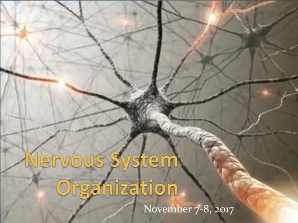
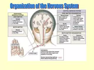
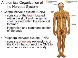
Nervous system organization. fig 6-37. Brain structure. fig 6-38. Brain structure (notes). Cerebrum structure Structure lobes: frontal, parietal, occipital, temporal (named for bones) cerebral hemispheres, subcortical nuclei (basal ganglia) Functions

E N D
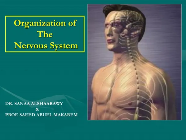
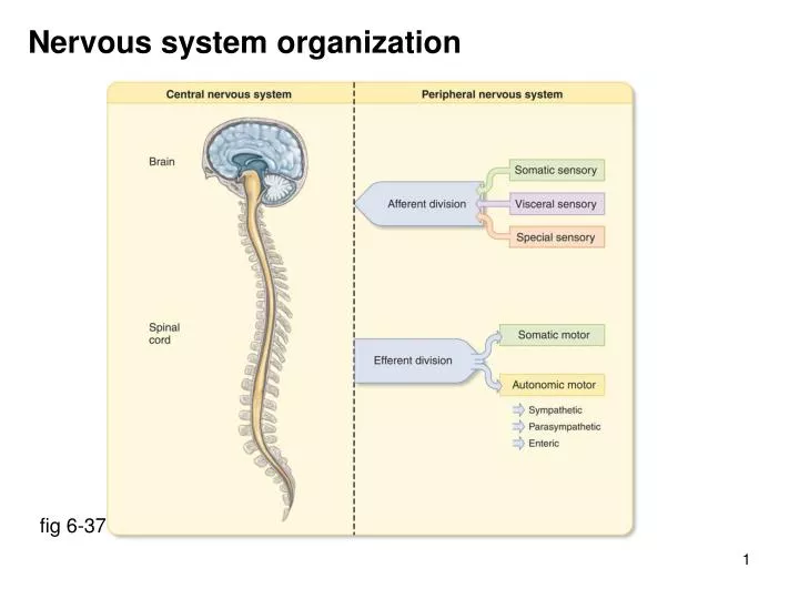
Nervous system organization fig 6-37
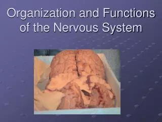
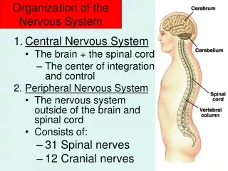
Brain structure fig 6-38
Brain structure (notes) Cerebrum structure Structure lobes: frontal, parietal, occipital, temporal (named for bones) cerebral hemispheres, subcortical nuclei (basal ganglia) Functions motor functions, sensory functions learning, reasoning, memory Thalamus (paired) Hypothalamus (paired) Cerebellum Brain stem = midbrain + pons + medulla oblongata
Cerebral hemispheres: motor function Primary & supplementary motor cortex (frontal lobe) skilled movements, particularly of distal muscles fig 10-10
Cerebral hemispheres: motor function Primary motor cortex motor “homunculus”; area related to fine motor control fig 10-11
Cerebral hemispheres: basal nuclei Basal nuclei coordination of motor activity, control movement & posture defective in Parkinson’s disease Corpus callosum: connection between right & left hemispheres fig 6-39
Cerebral hemispheres: sensory function Visual cortex (occipital lobe) Auditory cortex (temporal lobe) Taste cortex (parietal lobe) fig 7-14
Cerebral hemispheres: sensory function Somatosensory cortex (parietal lobe) touch, pressure, kinesthesia, temperature, pain sensory “homunculus” fig 7-20
Cerebral hemispheres: sensory function Olfactory cortex (frontal lobe) only sense not via thalamus, ~1000 odorant receptors afferent fibers to olfactory cortex & limbic system fig 7-45
Cerebral hemispheres: learning & memory Declarative memory (language, names, faces, events, facts) Short term: involves hippocampus & other temporal lobe structures ongoing graded or action potentials susceptible to shock, trauma, coma, electroconvulsive therapy Long term: many areas of association cortex involves protein synthesis, synapses, post-synaptic receptors Procedural memory (actions, emotional responses, fears) Short term various parts of brain Long term basal nuclei, cerebellum, sensorimotor cortex
Thalamus Sensory relay (synapses) for all sensory input except olfactory fig 6-39 (cropped)
Hypothalamus Functions: regulates anterior pituitary gland (produces releasing factors) nuclei for posterior gland hormones (antidiuretic hormone & oxytocin) regulates thirst & hunger involved in regulation of autonomic system regulates reproductive system (hormones & autonomics) circadian rhythms (clock in suprachiasmatic nucleus) regulates body temperature (integrative center) important component of limbic system
Limbic system Components: thalamus, hypothalamus, hippocampus, septal nuclei, olfactory bulbs, cingulate gyrus Functions: emotional responses (subjective feeling & behavior), memory fig 6-40
Cerebellum Location: on brain stem, over pons Input from sensorimotor cortex and vestibular (balance), visual, & musculature Output to cerebral cortex & brainstem nuclei Functions: comparing intended movements with actual outcomes, and making appropriate adjustments; coordinates movements, some procedural memories Cerebellar disease: intention tremor
Brain stem Components: mid brain, pons, medulla oblongata General functions: ascending & descending pathways cranial nerve nuclei for III through XII brainstem nuclei associated with posture, balance & walking reticular activating system centers for control of cardiovascular, respiratory, swallowing, vomiting
Reticular activating system Location: loose network in brain stem, widely distributed input/output Function: arousal (positive feedback), wakefulness, awareness, focus function essential for life
Spinal cord Gray matter: cell bodies & synapses White matter: ascending & descending tracts Ventral root: efferent pathways Dorsal root: afferent pathways Dorsal root ganglion: cell bodies of afferent neurons fig 6-41
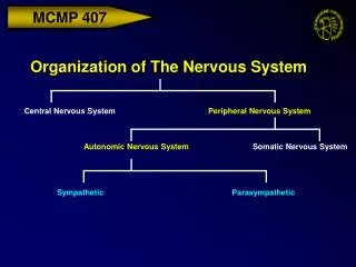
Nervous system organization fig 6-37
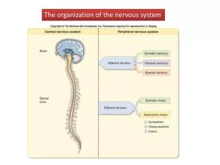
Peripheral nervous system: efferent division fig 6-43 Somatic: single neuron, innervates skeletal muscle, voluntary control Autonomic: 2 neuron chain, innervates smooth, cardiac muscle, glands, largely involuntary control
Autonomic nervous system: anatomy fig 6-46 cropped Parasympathetic: long preganglionic, short post ganglionic, ganglia often in end organ, little divergence, specific action Sympathetic: short perganglionic, long post ganglionic, much divergence adrenal medulla: endocrine supplement, acts as a unit
Autonomic nervous system: neurotransmitters * some exceptions adrenal medulla: preganglionic - acetyl choline postganglionic (chromaffin cell) - 80% epi, 20% norepi fig 6-46 cropped
Autonomic system distribution parasympathetic: craniosacral outflow long preganglionic especially vagus (X) sympathetic: thoracolumbar outflow short preganglionic fig 6-44
Autonomic nervous system: actions General: parasympathetic (PS): specific actions (rest, digest, rebuild) sympathetic (SY): general action (fight, fright, flight) Models: PS: “Slats Grobnik” lazy, overeating, overdrinking SY: “Og” caveman chased by saber toothed tiger
Autonomic nervous system: actions Heart: PS: rate SY: rate, contractility, cardiac output, blood pressure Vasculature: PS: minimal effects SY: arterioles constrict (especially skin, GI tract, kidneys), veins constrict Lungs: PS: bronchioles constrict, secretion SY: bronchioles dilate, secretion Gastrointestinal system: PS: secretions, motility, sphincters relax, blood to salivary glands SY: secretions, motility, sphincters constrict, blood to salivary glands
Autonomic nervous system: actions Metabolism: PS: anabolic ( insulin), synthesis of fat, glycogen, protein SY: catabolic ( epinephrine, glucagon, cortisol), lipolysis, glycogenolysis, gluconeogenesis Urinary bladder: PS: sphincter relaxes, bladder wall contracts (micturition), SY: sphincter constricts, bladder wall relaxes Sweat: PS: insignificant innervation (Vander is wrong!) SY: hands, feet (adrenergic), general (AcCh) – “cold sweat” Reproductive system: PS: erection (penis, clitoris) – NO is neurotransmitter, not AcCh SY: ejaculation (male), orgasm (female)
Adrenergic receptors Norepinephrine (NE) and epinephrine (E) bind to “adrenergic” receptors Examples: 1, 2, 1, 2, 3 NE & E bind to all types, although NE prefers ’s & E prefers ’s 1 receptors: cause smooth muscle contraction (e.g. arterioles, veins) have IP3, DAG, Ca++ as second messengers 1 receptors: are in (1) heart and mediate cardiac responses to SNS have cAMP as second messenger 2 receptors: cause relaxation of smooth muscle (e.g. (2) lung bronchioles) have cAMP as second messenger Smooth muscle response depends on receptor profile