Download

1 / 92
940 likes | 986 Views
DEVELOPMENT OF URINARY SYSTEM AND ANOMALIES. The urinary system consists of 1) Kidney 2) Ureter 3) Urinary bladder 4) Urethra. Urogenital system consists of urinary and genital systems.

E N D
The urinary system consists of 1) Kidney 2) Ureter 3) Urinary bladder 4) Urethra
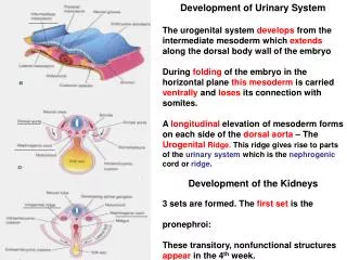
Urogenital system consists of urinary and genital systems. • It develops from; intermediate mesoderm, part of cloaca, coelomic epithelium covering the intermediate mesoderm. • Intermediate mesoderm is lying between somites and lateral plate mesoderm. The intermediate mesoderm produces a bulge called urogenital ridge.This ridge is divided into genital and nephrogenic ridges.
Nephrogenic cord Dorsal mesentery Gut Bulging on the posterior abdominal wall Covered by coelomic epithelium From cervical to sacral region
Cloaca –is the terminal dilated part of hind gut. • It divides into two parts by urorectal septum , primitive urogenital sinus and primitive rectum. • The primitive urogenital sinus further divides in to vesico urethral canal and definitive urogenital sinus. It further divides into pelvic phallic parts.
Derivatives of nephrogenic cord • Excretory tubules • mesonephric duct • Paramesonephric duct • gonad
Vertebrate kidney pass through 3 stages of evolution Three sets of kidneys develop in human embryo 1) pronephros 2) mesonephros 3) metanephros
The mesonephric kidney consists of 1) glomerulus 2) mesonephric tubule 3) mesonephric duct
Mesonephric tubules are formed successively in cranio caudal direction. • The cells of intermediate mesoderm first form the solid clusters of the cells. • Lumen appears in these clusters and converts them as vesicles. • These vesicles then become pear shaped. • Further elongation of pear shaped vesicles will form the mesonephric tubules.
First excretory tubules of mesonephros appear early in 4th week of development • Excretory tubules legnthen rapidly to form S-shaped loops • Acquire a tuft of capillaries at their medial extremity Around glomerulus tubules form Bowman’s capsule Together forming renal corpuscle Tubules enter longitudinal mesonephric or …….wolffian duct
Mesonephric kidney fails to perform the function of permanent kidney.
METANEPHROS • Appears in 5th week • The definitive human kidney consists of 1) collecting part ureteric bud 2) excretory part metanephricblastema 3) Angiogenicmesenchyme (The formative, undifferentiated material from which cells are formed)
Metanephros –The permanent kidney develops from two sources, one is the mesoderm of sacral part of nephrogenic cord ,and other is ureteric bud, which is a diverticulum arising from the mesonephric duct. • The ureteric bud arises from posteromedial aspect of mesonephric duct, just before it opens into the cloaca. The ureteric bud grows dorsally and reaches sacral part of nephrogenic cord .
The mesoderm of nephrogenic cord condenses around the tip of ureteric bud to form metanephric cap or metanephricblastema. • The contact of ureteric bud with metanephricblastema induces the division of ureteric bud. • The dilated terminal part of ureteric bud forms the pelvis of ureter and proximal part forms ureter itself.
The first three or four divisions of ureteric bud fuse to form the major calyces while next three to four divisions form minor calyces. • Further divisions of ureteric bud form the collecting tubules. • The mesodermal cells of metanephricblastema to form a cluster of cells around it. • The cluster acquire a lumen and form the vesicle.The vesicle elongate to form the tubules.
The distal end of the tubule is invaginated by capillaries to form the glomerulus • Further elongation of tubules will take place and various parts of nephrons are formed such as Bowmanscapsule,PCT,DCT loop of Henle. • Thus the secretary part of kidney(nephrons) is derived from metanephricblastema,while collecting part(collecting tubules,minor,majorcalyces,pelvis of ureter and ureter proper) is derived from ureteric bud.
Splits into cranial & caudal portions: Future major calyces
Each calyx forms 2 new buds while penetrating metanephric tissue • Buds continue to subdivide until 12 or more generations of tubules • Until 5th month tubules form • The branches of 2nd,3rd,4th orders are absorbed to form the minor calyces
All branches from the 5th and subsequent orders persists as the collecting tubules of the permanent kidney. • They converge on minor calyx forming renal pyramid
Derivatives of ureteric bud • Ureter • Renal pelvis • Major calyces • Minor calyces • 1-3million collecting tubules
EXCRETORY PART It is developed from the metanephric blastema
Each newly formed collecting tubule is covered at its distal end by metanephric tissue cap
Under inductive influence of tubule. Cells of tissue cap form renal vesicles • These vesicles are the precursors of the nephrons of the kidney
Continuous legnthening of excretory tubule results in formation of proximal convoluted tubule, loop of Henle & distal convoluted tubule
Summary • Beginning of 4th week: pronephros: 7-10 cell groups • End of 4th week: pronephric system disappear • Early in 4th week: excretory tubules of mesonephros appear • Middle of 2nd month: mesonephros is large ovoid organ • End of 2nd month: mesonephric tubules degenerate mostly • 5th week: metanephros appear
Nephrons are formed until birth • At birth approximately 1 million in each kidney • The tubular function comes out at about the 9th week • Differentiation of glomerular capillaries start from 10th week • Urine production follows soon. • Definitive kidney formed from metanephros becomes functional near 12th week • Reabsorption of filtrate by the loop of Henle takes place at about the 13th week
Kidney have lobulated appearance at birth • Lobulation disappears during infancy as a result of further growth of nephron although there is no increase in their number
FACTORS RESPONSIBLE FOR THE ASCENT • Continuous lengthening of the ureteric bud • Diminution of the fetal curvature • Growth of body in lumbar and sacral region • Too small pelvic cavity for the accommodation of the renal growth
ANOMALIES OF THE KIDNEYanomalies of position • Ectopic kidney
ANOMALIES OF THE KIDNEY • Duplication of urinary tract
ANOMALIES OF THE KIDNEY • Agenesis • Hypoplasia/ Hyperplasia- underdeveloped/overdeveloped • Hydronephrosis- distension of pelvis • Duplication- extra kidney, separate or fused to normal kidney
Pelvic kidney Failure of kidney to ascend through arterial fork
ANOMALIES OF THE KIDNEYanomalies of shape • Horse shoe kidney- lower poles fused • Pancake kidney- 2 kidneys as 1 mass • Lobulated kidney- fetal lobulation persists
Horseshoe kidney Kidneys pushed together during passage through arterial fork Lower lumber Ascent prevented by inferior mesenteric artery
ANOMALIES OF THE KIDNEY • Malrotated kidney: hilum directed forwards (non rotation),anteromedially(incomplete rotation), or anterolaterally(reverse rotation), • Congenital polycystic kidney • Accessory renal artery: persistence of embryonic vessels formed during ascent of kidney • Tumours of kidney: Wilm’stumour WAGR syndrome: aniridia, hemihypertrophy, Wilm’stumour