Download

1 / 66
730 likes | 776 Views
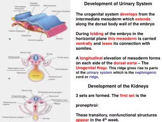
Development Of Urinary System. Dr Mukesh Singla Additional Professor, Anatomy AIIMS, Rishikesh. Intermediate Mesoderm Forms Much of the Urogenital System. Mesoderm in the Chick Embryo. Lateral Rotation. Hindgut with allantois. Hind Gut-Cloaca. Urorectal Septum. Urorectal Septum.

E N D
Development Of Urinary System Dr MukeshSingla Additional Professor, Anatomy AIIMS, Rishikesh
The Ureteric Bud Induces the MetanephricBlastema to Form Filtration Units
MCQ When does the metanephros become functional? (A) At week 3 of development (B) At week 4 of development (C) At week 10 of development (D) Just before birth (E) Just after birth
Renal Tumors and Defects • Wilms' tumor is a cancer of the kidneys that usually affects children by 5 years of age but may also occur in the fetus. • Wilms' tumor is due to mutations in the WT1 gene on 11p13, and it may be associated with other abnormalities and syndromes. • For example, WAGR syndrome is characterized by aniridia, Genitourinary abnormalities, and Wilms' tumor and mental retardation • Denys-Drash syndrome consists of renal failure, pseudohermaphrodism, and Wilms' tumor.
Renal dysplasias and agenesis • Multicystic dysplastic kidney in which numerous ducts are surrounded by undifferentiated cells. • Nephrons fail to develop • ureteric bud fails to branch, • the collecting ducts never form. In some cases, these defects cause involution of the kidneys and renal agenesis
Position of the Kidney • The kidney, initially in the pelvic region, later shifts to a more cranial position in the abdomen. This ascent of the kidney is caused by • diminution of body curvature • by growth of the body in the lumbar and sacral regions In the pelvis, the metanephros receives its arterial supply from a pelvic branch of the aorta. During its ascent to the abdominal level, it is vascularized by arteries that originate from the aorta at continuously higher levels. The lower vessels usually degenerate, but some may remain.
Developmental anomalies of the kidney A. Renal agenesis : • Failure of one or both kidneys to develop • Bilateral: rare, associated with other congenital anomalies, incompatible with life • Unilateral: common (one in 4,000 infants, more boys than girls) , asymptomatic; • Other kidney enlarges to compensate#Absentipsilateral renal artery
In congenital polycystic kidney disease • It may be inherited as an autosomal recessive or autosomal dominant disorder or may be caused by other factors. Autosomal recessive polycystic kidney disease • which occurs in 1/5,000 births, is a progressive disorder in which cysts form from collecting ducts. The kidneys become very large, and renal failure occurs in infancy or childhood. In autosomal dominant polycystic kidney disease, cysts form from all segments of the nephron and usually do not cause renal failure until adulthood. The autosomal dominant disease is more common (1/500 to 1/1,000 births) but less progressive than the autosomal recessive disease.
Polycystic Kidney Recessive form: 1:5000; cysts from collecting tubules; renal failure in infancy Dominant form: 1:500-1000: cysts from anywhere; renal failure in adulthood
Duplication of the ureter • results from early splitting of the ureteric bud • Splitting may be partial or complete, and metanephric tissue may be divided into two parts, each with its own renal pelvis and ureter. More frequently, however, the two parts have a number of lobes in common as a result of intermingling of collecting tubules. • In rare cases, one ureter opens into the bladder, and the other is ectopic, entering the vagina, urethra, or vestibule .This abnormality results from development of two ureteric buds. One of the buds usually has a normal position, whereas the abnormal bud moves down together with the mesonephric duct. Thus it has a low, abnormal entrance in the bladder, urethra, vagina, or epididymal region.
MCQ During surgery for a benign cyst on the kidney, the surgeon notes that the patient’s right kidney has two ureters and two renal pelves. This malformation is • (A) an abnormal division of the pronephros • (B) an abnormal division of the mesonephros • (C) formation of an extra mass of intermediate mesoderm • (D) a premature division of the metanephric blastema • (E) a premature division of the ureteric bud
MCQ • The transitional epithelium lining the urinary bladder is derived from • (A) ectoderm • (B) endoderm • (C) mesoderm • (D) endoderm and mesoderm • (E) neural crest cells