Download
1 / 45
450 likes | 1.29k Views
Nasopharyngeal tumours. ENT Department Registrar FRCS Teaching Freeman Hospital Newcastle April 2010 Cameron Davies-Husband MBBS, BSc (Hons), MRCS (Eng), DOHNS (Eng), PGCert. Aims. To provide an overview of nasopharyngeal carcinoma, including: Pathology Symptoms Diagnosis Treatment.

E N D
Nasopharyngeal tumours ENT Department Registrar FRCS Teaching Freeman Hospital Newcastle April 2010 Cameron Davies-Husband MBBS, BSc (Hons), MRCS (Eng), DOHNS (Eng), PGCert
Aims • To provide an overview of nasopharyngeal carcinoma, including: • Pathology • Symptoms • Diagnosis • Treatment
Introduction • Nasopharyngeal carcinoma: • non-lymphomatous, squamous-cell carcinoma occurring in the epithelial lining of the nasopharynx.
Relevent anatomy • Nasopharynx is located below the central skull base • The sphenoid sinus forms the roof • The roof slopes posteriorly with the basiocciput and upper cervical spine forming the posterior wall • The lateral nasopharynx wall is formed by: • the medial pterygoid plate, • the palatal muscles, • the torus tubarius • the fossa of Rosenmuller • The soft palate separates the nasopharynx from the oropharynx. • The fossa of Rosenmuller is located superior and posterior to the torus tubarius
Modes of spread • Anterior: • Infiltration of pterygopalatine fossa through the sphenopalatine foramen9 • Earliest sign of involvement of the pterygopalatine fossa is obliteration of the normal fat content
Modes of spread • Anterior: • Perineural spread along the mandibular nerve can seen to extend into the intracranial cavity through the foramen rotundum • The tumour can infiltrate further superiorly into the inferior orbital fissure and subsequently extend into the orbital apex
Modes of spread • Lateral: • Partial or complete effacement of the fat-filled parapharyngeal space • The mandibular nerve is often infiltrated. • Posterolateral spread into the retrostyloid compartment may result in the infiltration of the carotid space placing cranial nerves IX, X, XI and XII at risk12,13.
Epidemiology • Uncommon: age-adjusted incidence for both sexes is < 1 per 100, 000 population1 • Occurs with much greater frequency in: • Southern China • Northern Africa • Alaska • Incidence among men and women in Hong Kong is 20–30 and 15–20 per 100 000, respectively1 • Lower among Chinese people born in North America7,8
Pathology • Tumour cells are of squamous origin10 • The undifferentiated carcinoma is a form of squamous-cell carcinoma11 • EBV-encoded RNA signal has been shown, by in-situ hybridisation, to be present in nearly all tumour cells12 • Premalignant lesions also harbour EBV12
Pathology • Histological classification; WHO (1978)15: • Type I: typical keratinising squamous-cell carcinomas • Type II: non-keratinising squamous carcinomas • Type III: undifferentiated carcinomas • The second classification is correlated with EBV serology: • Squamous-cell NPCs have reduced EBV titre • Undifferentiated NPCs have raised EBV titres.
Pathology • In North America: • 25% of tumour patients have type I histology • 12% have type II • 63% have type III. • In Southern China: • 2% of tumour patients have type I histology • 3% have type II • 95% have type III17
Classification • Keratinising squamous-cell carcinoma (WHO type I) • Non-keratinising carcinoma • Differentiated non-keratinising carcinoma (WHO type II) • Undifferentiated carcinoma (WHO type III)
Symptoms • (1) presence of tumour mass in the nasopharynx • (2) dysfunction of the eustachian tube, associated with the lateroposterior extension of the tumour to the paranasopharyngeal space • (3) skull-base erosion and palsy of the fifth and sixth cranial nerves, associated with the superior extension of the tumour • (4) neck masses, usually appearing first in the upper neck, associated with metastases
Symptoms • A retrospective analysis of 4768 patients identified symptoms of nasopharyngeal carcinoma at presentation21: • Neck mass (76%) • Nasal dysfunction (73%) • Aural dysfunction (62%) • Headache (35%) • Diplopia (11%), • Facial numbness (8%) • Weight loss (7%) • Trismus (3%)
Symptoms • Physical signs present at diagnosis included21,22: • Enlarged neck node (75%) • Cranial nerve palsy (20%) • 3rd, 5th, 6th, and 12th nerves • Investigations: • EBV serology • EUA & PNS biopsy • Cross sectional CT/MRI
Imaging studies • Cross sectional CT: • Paranasopharyngeal spread: one of the most common modes of NPC extension28 • Perineural spread (through the foramen ovale) • Important route of intracranial extension28 • Cavernous sinus involvement without skull-base erosion29
Imaging studies • MRI > CT for displaying both superficial and deep nasopharyngeal soft tissue and for differentiating tumour from soft tissue. • MRI is also more sensitive for assessment of retropharyngeal and deep cervical nodal metastases30 • CT should be undertaken when the status of the skull base cannot be satisfactorily established with MRI31 • MRI is able to detect marrow infiltration by tumours, whereas CT cannot (associated with an increased risk of distant metastases32)
Imaging studies • Distant metastases: • Bone scans33 liver scintigraphy34 abdominal ultrasonography35 and marrow biopsy36 are of little valueinroutine staging • No evidence to lend support to distant imaging for low-risk (N0 or stage I) disease • High-risk (N3) disease should be fully staged with chest radiography, bone scan, and liver ultrasonography37 • The role of PET scanning is established, but it’s value has not been ascertained38
Imaging studies • Monitoring post treatment: • Both CT and MRI have: • Low sensitivity41 • Moderate specificity41 • MRI generally better at detecting tumour recurrence and post radiotherapy complications42 • PET reported to be more sensitive then CT/MRI at detecting recurrent/residual tumours43
Staging • American Joint Committee on Cancer Staging and End Result Reporting/International Union Against Cancer (AJC/UICC) • Preferred staging system in USA/Europe • Revised in 1997: • Enabled patients to be staged more sensitively55 • Provided a better predictor of survival than the old system56
Staging • T1 + T2 new “T1” tumours ~ confined to nasopharynx • T2 tumours ~ involving nasal fossa, oropharynx, or paranasopharyngeal space • T3 tumours ~ extension to the skull base/paranasal sinuses • T4 tumours ~ extended into the infratemporal fossa, orbit, hypopharynx, and cranium, or to the cranial nerves
Staging • N1: unilateral nodal involvement • N2: bilateral nodal disease that has not reached N3 designation, irrespective of the size, number, and anatomical location of the nodes • N3: lymph nodes larger than 6cm (N3a), or nodes that have extended to the supraclavicular fossa (N3b)54
Prognosis • Tumour extent ~ TNM classification • Size/degree of fixation of lymph nodes57 • Patient gender57 • Patient age57 • The presence of cranial nerve palsy57 • Ear symptoms at presentation57 • Histologicaltype58 • Radiotherapy dose and coverage58 • Paranasopharyngeal extension59
Prognosis • Estimated 1% increase in risk of local failure for every 1 cm3 increase in primary tumour volume63 • Quantitative analysis of circulating EBV DNA in NPC positively correlates with disease stage, as well as exhibiting prognostic importance64
Prognosis • T1–2 N0–1 (relatively good treatment outcome) • T3–4 N0–1 (mainly local failure) • T1–2 N2–3 (mainly regional and distant failure) • T3–4 N2–3 (local, regional and distant failure) • Early evidence that for advanced diseases, adding chemotherapy to radiotherapy will improve treatment outcome, both in terms of locoregional control and distant metastases66,67
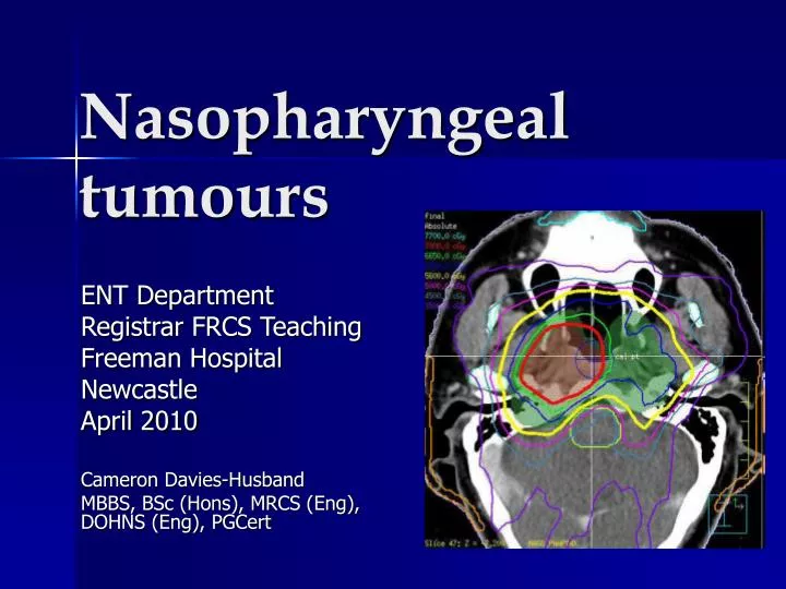
Treatment • Radiotherapy: the standard treatment for nasopharyngeal carcinoma • Dose-limiting organs at risk: • brain stem, spinal cord, pituitary-hypothalamic axis, temporal lobes, eyes, middle and inner ears, and parotid glands
Treatment • Radiotherapy approaches: • Phase I treatment: • Large lateral opposing faciocervical fields that cover the primary tumour and the upper neck lymphatics in one volume • Matching lower anterior cervical field for lower neck lymphatics • When the spinal cord dose reaches 40–45 Gy phase II treatment
Treatment • Phase II treatment – either: • Lateral opposing facial fields with anterior facial field for the primary tumour, with matching anterior cervical field • Lateral opposing facio-cervical fields but with shrinkage of fields to avoid the spinal cord68,69
Treatment • Radiotherapy tumour control: • T1-T2: 75–90% • T3-T4: 50–75%65,68,69,70,71 • Radiotherapy nodal disease control: • N0-N1: 90% • N2-N3: 70%65 • Note: high incidence of occult neck node involvement - prophylactic neck radiation is usually recommended73
Treatment • Good locoregional control should be the prime objective of treatment • Locoregional relapses represent a significant risk factor for the development of distant metastases74 • For T1-T2 tumours, a booster dose via intracavitary brachytherapy improved tumour control by 16%75
Treatment • Intensity modulated radiotherapy (IMRT) • Overcomes major limitations of 2D planning78,79 • Improves the dose differential between the tumour and the dose-limiting organs81,82 • Resolves the problem of dose uncertainty at the junction between the primary tumour and neck lymphatic target volumes as it enables the primary tumour and the upper neck nodes to be treated in one volume throughout • Excellent locoregional control of nasopharyngeal carcinomas83
Chemotherapy • Twelve RCT’s have reported on neoadjuvant, concurrent, and adjuvant therapy, or on a combination of these approaches. • Nine were reported before 2004 and included four neoadjuvant chemotherapy studies,91–94 three concurrent chemotherapy studies,66,67,95 and two adjuvant studies96,97 • One of the concurrent studies95 has recently been updated,98 and two of the neoadjuvant studies92,93 have been updated and pooled for meta-analysis99 • Three more concurrent chemotherapy studies have been reported from Hong Kong and Singapore100–102
Chemotherapy • Meta-analysis99 • Improvements in relapse-free survival and disease specific survival • Overall survival was not improved • Adjuvant studies96,97: • No improvement either in relapse-free survival or overall survival • Of the three basic approaches tested (neoadjuvant, concurrent, and adjuvant chemotherapy), concurrent chemoradiotherapy is the most efficacious…
Follow-up • Confirmation of disease remission: • Clinical examination • Endoscopic examination (+/- biopsy) • Imaging studies • Residual tumours can be treated with either: • Cone down fields111 • Brachytherapy112 • Residual neck node disease is amenable to radical neck dissection.
Follow-up • Regular imaging every 4–6 months during the initial 3–5 years after treatment is advised 114,115 • Endoscopic examination to detect superficial tumours • Cross-section imaging to detect deep infiltrating tumours not associated with mucosal lesion31 • PET superior to MRI for detection of residual and recurrent tumour46
Follow-up • Circulating free EBV DNA has been reported in patients with nasopharyngeal carcinoma117 • Quantities of EBV DNA copies before and after treatment are significantly related to the rates of overall and disease-free survival119 • Serum EBV DNA shown to be more sensitive and reliable than other options for the detection of distant metastases116
Follow-up • Levels of post-treatment EBV DNA compared with pre-treatment EBV DNA are a good predictor of progression-free survival120 • Raised EBV DNA detected in only 67% of patients with locoregional recurrence,116,122 • In those with distant metastasis levels of EBV DNA copies were heightened before the appearance of clinical abnormality116
Management of residual/recurrent disease • Notoriously difficult to confirm because only clusters of tumour cells are present140 • Survival after re-treatment for more extensive disease remains poor, but is still higher than in patients receiving supportive treatment only113
Recurrent disease in the neck • After combined chemoradiation for nasopharyngeal carcinoma, isolated failure in the neck is less than 5%146 • Further course of external radiotherapy: the overall 5-year survival rate is around 20%147 • Radical neck dissection: 5-year tumour control rate of 66% in the neck and a 5-year survival of 38%142 • Radical neck dissection PLUS brachytherapy if tumour spreads beyond lymph node148
Recurrent disease in the nasopharynx • Can be managed with a second course of external radiotherapy • The dosage should be greater than the initial radiation dose • Incidence of late sequelae after re-irradiation is 24% with treatment mortality of 1·8%149 • Small localised tumours: • Stereotactic radiotherapy (2-year local tumour control rate of • 72%77) • Brachytherapy (5-year local tumour control rates were 87% and 63% for persistent/recurrent tumours respectively) • Surgical resection
Recurrent disease in the nasopharynx • Extensive disease • Nasopharyngectomy • 5-year actuarial control of tumours in the nasopharynx is about 65% • 5-year disease-free survival rate is around 54%.159,160
New developments • Gene therapy: • Replication-deficient adenovirus vector; transgene expression is under the transcriptional regulation of oriP of EBV has been reported174 • Immune therapy: • Augmentation of cytotoxic T-lymphocyte responses175 • Adoptive transfer of autologous EBV-specific cytotoxic T-cells176


![Nasopharyngeal Cancer [6]](https://cdn1.slideserve.com/3354426/nasopharyngeal-cancer-6-dt.jpg)