Download
1 / 68
680 likes | 699 Views
Chronoamperometric studies of the adhesion-spreading events of lipid vesicles on electrodes. Fritz Scholz Victor Agmo Hernández, Dirk Hellberg, Michael Hermes, Andreas Gröning, Stefan Zander University of Greifswald, Institute of Biochemistry Greifswald, Germany.

E N D
Chronoamperometric studies of the adhesion-spreading events of lipid vesicles on electrodes Fritz Scholz Victor Agmo Hernández, Dirk Hellberg, Michael Hermes, Andreas Gröning, Stefan Zander University of Greifswald, Institute of Biochemistry Greifswald, Germany
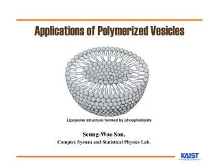
Cylindrical micelle Self-assembled lipid structures Micelle DMPC = Dimiristic-phosphatidyl- choline Liposome
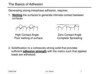
Hydrophilic head Hydrophobic tail
Hydration water = water dipoles interact with polar headgroup Water having ‘iceberg structure’ = water dipoles do not interact with the hydrophobic tail; they loose conformational entropy(about RT ln2 = 0.42 kcal /mol for each water molecule)
H H H H H H O O O O O O H O H H H H H H H hydrophobic surface H H H H H H H H H O O O O O O O O O H H H H H H H H H Iceberg water:3 possible configurations for each water tetrahedron Entropy gain Entropy loss Liquid water:6 possible configurations for each water tetrahedron
The hydrophobic interaction is mostly an entropic effect originating from the disruption of highly dynamic hydrogen bonds between molecules of liquid water by the nonpolar solute. A hydrocarbon chain or a similar nonpolar region or a big molecule is incapable of forming hydrogen bonds with water. Introduction of such a non-hydrogen bonding surface into water causes disruption of the hydrogen bonding network between water molecules. The hydrogen bonds are reoriented tangentially to such surface to minimize disruption of the hydrogen bonded 3D network of water molecules and thus leads to a structured water "cage" around the nonpolar surface. The water molecules that form the "cage" (or solvation shell) have restricted mobilities. In the solvation shell of small nonpolar particles, the restriction amounts to some 10%, e.g. in the case of dissolved Xe at room temperature, a mobility restriction of 30% has been found. In the case of larger nonpolar molecules the reorientational and translational motion of the water molecules in the solvation shell may be restricted by a factor of two to four. Thus at 25°C the reorientational correlation time of water increases from 2 to 4-8 picoseconds. Generally, this leads to significant losses in translational and rotational entropy of water molecules and makes the process unfavorable in terms of free energy of the system. By aggregating together, nonpolar molecules reduce the surface area exposed to water and minimize their disruptive effect.
All iceberg water is released = enormous entropy gain! The driving force of bilayer formation is the entropy gain behind the so-called hydrophobic interaction (there is also a small energy gain due to dispersion interaction between alkyl chains: about 6.9 kJ/mol per CH2 group for alkyl chains in a hexagonal monolayer)
The Hydrophobic Effect: Protein folding Minimizing the number of hydrophobic side-chains exposed to water is an important driving force behind the folding process. Formation of intramolecular hydrogen bonds provides another important contribution to protein stability.
The Hydrophobic Effect: Formation of micelles, liposomes, membranes
The Hydrophobic Effect: Immiscibility of water and oil Dissolution of a hydrocarbon in water would need the formation of ordered water shells around the single molecules. As this is entropically discouraged, the hydrocarbon will instead form an own phase to minimize the contact surface area with water.
The Hydrophobic Effect: Hydrophobic surfaces are not (or only badly) wetted by water
What happens when a liposome approaches a mercury electrode? Hg ?
“Bioparticles” Lecithin liposomesThrombocyte vesicles Electrochem. Commun. 4 (2002) 305-309.. Bioelectrochem. 74 (2008) 210-216. J. Phys. Chem. B 109 (2005) 14715-14726. Langmuir 22 (2006) 10723-10731. Israel J. Chem. 48 (2008) 169-184. J. Solid State Electrochem. 13 (2009) 639-649. J. Solid State Electrochem. 13 (2009) 1111-1114. Bioelectrochem. 74 (2008) 149-156. Mitochondria Cardiolipin liposomes Angew. Chem. 123 (2011) 7004 –7007 in preparation Angew. Chem. Int. Ed. 50 (2011) 6872 –6875 Reconstituted plant plasma membrane vesicles submitted “Non-bioparticles” Clay (montmirollonite) particlesElectrochem. Commun. 6 (2004) 929-933 Silver nanoparticles, etc.: Richard Compton’s group
What happens when a liposome approaches a mercury electrode? Hg ?
This process produces capacitive current current peaks under potentiostatic control -0.2 V vs. Ag|AgCl, 25 °C -0.7 V vs. Ag|AgCl, 25 °C pzc (-0.45 V vs. Ag|AgCl) Hellberg, D.; Scholz, F.; Schauer, F.; Weitschies, W.; Electrochem. Commun.2002, 4, 305
DMPC GUVs, 0.1 g L-1 in 0.1 M KCl. Potential: -0.9 V vs. Ag|AgCl The overall kinetics • Macroscopic: frequency of detectable events
Adhesion-spreading of one single liposome Current sampling each 50 µs (20 kHz sampling)
Data about the liposome • Area of the electrode covered by the lecithin molecules • Number of lecithin molecules forming the liposome Qlip= Aisland· (qelectrolyte - qmonolayer)
Size distribution • If unilamellar spherical vesicles are assumed:
Size distribution and lamellarity • If multilamellar, compact spherical vesicles are assumed:
Microscopical kinetics of adhesion-spreading • Integrated peak Chi2 R2 ---------------------------------------- 4.9747×10⁻²⁷ 0.99994 ---------------------------------------- Hellberg, D.; Scholz, F.; Schubert, F.; Lovrić, M.; Omanović, D.; Agmo Hernández, V.; Thede, R.; J. Phys. Chem. B2005, 109, 14715
1st step: the liposome interacts with the electrode surface (interaction-docking) Hg
Study of the adhesion-spreading of a single liposome Theoretical model Hellberg, D.; Scholz, F.; Schubert, F.; Lovrić, M.; Omanović, D.; Agmo Hernández, V.; Thede, R.; J. Phys. Chem. B2005, 109, 14715
Activation energies For DMPC GUVs in 0.1 M KCl detected at -0.9 V vs. Ag|AgCl Bilayer opening Rupture-spreading Ea(opening): 34.7 ± 5 kJ mol-1 Ea(opening): 49.6 ± 2.2 kJ mol-1 Ea(spreading): -9 ± 4.9 kJ mol-1 Ea(spreading): 33.8 ± 11.5 kJ mol-1
Dependence of the time constants on the vesicle lamellarity and size • In the liquid crystalline phase (40 º C) t1 standard deviation MLVs: 77% GUVs: 37% t2 standard deviation MLVs: 41% GUVs: 18% Agmo Hernández, V.; Scholz, F.; Langmuir2006, 22, 10723
Rupture-spreading Agmo Hernández, V.; Scholz, F.; Langmuir2006, 22, 10723 Dependence of the time constants on the vesicle size • In the rippled gel phase (13 ºC)
Dependence of the activation parameters on the vesicle size • Rupture-spreading
Why a size dependence of the kinetic parameters? • Rupture-spreading - - - - - - - - - - - - - - + + + + + + + + + + + + + + + + + + + + + + + + + + + + + + + + + + + + + + + + + + + + + + + + + Hg
Pore formation • Energy barrier to overcome to build a pore The larger the liposome, the larger the energy barrier Needham, D.; Evans, E.; Biochemistry1988, 27, 8261 Lee, C.; Lin, W.; Wang, J.; Opt. Eng.2001, 40, 2077 Rawicz, W.; Olbrich, K. C.; McIntosh, T.; Needham, D.; Evans, E.; Biophys. J.2000, 79, 328
Dependence of the activation parameters on the vesicle size: Bilayer opening • Just the small liposomes have a clearly higher activation energy of bilayer opening
Interaction at the area of contact Turning around of lecithin molecules must take place Hg
Activated state of bilayer opening • Molecules in the process of turning around • Similar activated state like in flip-flop translocation • Flip-flop translocation activation energy: 81 kJ mol-1 [1] • Bilayer opening activation energy (Qlip = 0): 93 ± 19kJ mol-1 • [1] Kornberg, R. D.; McConnell, H. M.; Biochemistry1971, 10, 1111
Study of several membrane systems • MLVs with cholesterol • Effect of lytic peptides (mastoparan X, melittin) • Effect of surfactants (Triton X-100)
Phase diagram DMPC-Cholesterol Superlattices at certain cholesterol molar concentrations: 0.154, 0.2, 0.25, 0.33, 0.4
Activation energies at low temperatures Overall Bilayer opening Spreading V.Agmo Hernández, F.Scholz: Bioelectrochem. 74 (2008) 149-156
Activation energies at high temperatures Overall Bilayer opening Spreading V.Agmo Hernández, F.Scholz: Bioelectrochem. 74 (2008) 149-156
0 mM MPX 0.01 mM MPX 0.1 mM MPX Effect of lytic peptides • Adding Mastoparan X to GUVs in the gel phase: Reduction of the activation energy of rupture-spreading with 0.1 mM MPX: 30 kJ/mol V.Agmo Hernández, F.Scholz: Bioelectrochem. 74 (2008) 149-156
Effect of lytic peptides V.Agmo Hernández, F.Scholz: Bioelectrochem. 74 (2008) 149-156
Effect of surfactants V.Agmo Hernández, F.Scholz: Bioelectrochem. 74 (2008) 149-156
DMPC GUVs adhesion-spreading at 35 ºC Current sampling each 1.333 μs High resolution measurements
High resolution measurements Time constant for docking in the order of 5-15 ms!!
Activation energy of interaction-docking One and the same activation energy for both the gel and the liquid crystalline phases
Why? • The process represents the contact between the two double layers (first interaction)