Download

1 / 39
520 likes | 1.11k Views
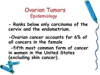
Benign Ovarian Tumors. Dr.Pravin Mhatre Associate Professor and Unit incharge, Nowrosjee Wadia Maternity Hospital and G.S. Medical College, Parel, Mumbai. Solid Ovarian Tumors. Child Germ cell Post pub. Germ cell , Dysgerminoma 20-35 Germ cel l, Dysgerminoma

E N D
Dr.Pravin Mhatre Associate Professor and Unit incharge, Nowrosjee Wadia Maternity Hospital and G.S. Medical College, Parel, Mumbai
Solid Ovarian Tumors • Child Germ cell • Post pub. Germ cell,Dysgerminoma • 20-35 Germ cell, Dysgerminoma • 35-45 Thecoma,Metastatic • Post meno. Thecoma,Metastatic Fibroma
Solid Ovarian Tumors • SMALL • Brenner • Fibroma • Theca cell tumor • Arrhenoblastoma • Dysgerminoma
Solid Ovarian Tumors • MEDIUM SIZE • Brenner • Fibroma (Capsule) • Granulosa cell tumor • Teratoma • Dysgerminoma (Capsule) • Sarcoma
Solid Ovarian Tumors • LARGE SIZE • Brenner • Fibroma (Capsule-Pedicle) • Krukenberg (Capsule-Pedicle) • Carcinomata
Cystic Ovarian Tumors • Child Terratoma, Granulosa cell • Post pub. Terrato, FOLL. • 20-35 Terrato,FOLL.Epith(B/M) • 35-45 Terrato,FOLL. • Post meno. Epith(B/M)
Cystic Ovarian Tumors • SMALL SIZE • Follicular cyst • Follicular hematoma • Corpus Luteual cyst • Corpus Luteual hematoma • Dermoid (ped. Capsulated)
Cystic Ovarian Tumors • MEDIUM SIZE • Cystoma simplex • Granulosa &Theca leutin cyst • Papillary serous cystadenoma • Granulosa • Dermoid (ped. Capsulated)
Cystic Ovarian Tumors • LARGE SIZE • Mucinous cystadenoma • Serous cystadenoma
Pedunculated Ovarian Tumors • Child Teratoma • Post pub. Teratoma • 20-35 Teratoma, Mucinous • 35-45 Teratoma,Mucinous • Post meno.Krukenberg (long pedicle) Fibroma
Capsulated Ovarian Tumors • Child Terato, Granulosa cell. • Post pub. Terato, GR. • 20-35 Terato, GR.,Mucin/Serous • 35-45 Terato, GR.,Mucin/serous • Post meno. GR.,Krukenberg, Sertoli.
Unilateral Ovarian Tumors • Child Terratoma • Post pub. Terrato, FOLL. • 20-35 Terrato,FOLL.Epith(B) Germ cell • 35-45 Terrato,FOLL.Epith(B) Germ cell • Post meno. Epith(B/M)
Bilateral Ovarian Tumors • Child Terratoma • Post pub. Terrato, • 20-35 Terrato,Epith(B) • 35-45 Terrato,Epith(B) • Post meno. Epith(B/M)
Ovarian Tumors • Ascites and Metastesis • Child • Post pub. Germ cell (M) • 20-35 Epith(M) • 35-45 Epith(M) • Post meno. Epith(M)
Ovarian Tumors Associations • Mucinous + Fibroma • Mucinous + Terratoma (same/opp) • Mucinous + Brenner
Ovarian Tumors Associations • Brenner + Terratoma Granulosa cell tumor Mucinous cystadenoma
Ovarian Tumors Associations • Granulosa cell tumor + Brenner Fibroma Terratoma Fibroid Endometrial hyperplasia Hyperthecosis
Ovarian Tumors Associations • Dermoid (Benign cystic terratoma) Granulosa cell tumor Thecoma Androblastoma
SPECIMENS • Dermoid • Mucinous cystadenoma • Meigs syndrome • Dysgerminoma • Granulosa cell Tumor • Arrhenoblastoma • Theca cell Tumor • Brenner • Krukenbergh • fibroma
Dermoid ( Benign cystic terratoma ) • 10% of all cystic swellings • 12% bilateral • Pedunculated • Well capsulated • 15 cm • Common on Right side • Multicentric
Dermoid ( Benign cystic terratoma ) • Ectoderm + Mesoderm + Endoderm • Supradiaphramatic structures • Teeth permanent never deciduous • Teeth canines,molars,incisors but never premolars • No Gonadal tissue • But assoc. with Granulosa Theca Androblastoma
Dermoid ( Benign cystic terratoma ) Complications • Torsion 10% • Rupture 1% • Infection • Hemolytic anemia-splenomegaly • Malignancy
Dermoid ( Benign cystic terratoma ) Radiological Signs • Presence of teeth and bone • Halo sign due to thick capsule • Calcification in capsule • Commonest tumor _ pregnancy
Dermoid ( Benign cystic terratoma ) • Dale’s sign • Kustner’s sign
Mucinous Cystadenoma • 10% Bilat, 6% malignant • Pedicle & large size = Complications • Pseudomyxoma Peritonei -2-5% • Mucin- ppt by alcohol • Pesudomucin-sol in water & dil acid - stained by Hematoxyline. - high con of neutral polysaccharide. • Silomucin res. to sialase = Malign. • Enzymes-Hyal, tryp, amyl, pep, peroxidase
Meig’s Syndrome • Demons (Fra) & Lawson Tait (Briton) • Criteria for Meig’s Syndrome (3) • Psuedo-Meig’s • Theca/Granulosa Tumor • Hyperstimulaion • Ovarian malig. • Struma ovari 10% • Serous/Mucinous
Dysgerminoma • 1906-Chevassu Dis = neutral/disgermal • 1911-Chenot Dys = primitive germ cell yolk sac – gonadal ridge same sex as host • Japan &India high incidence • Age 20 • 50% unilateral Rt sided, 20% bilateral • Size 3-50 cm ,common size 15cm. • Intersex-xo,xy,xxx Fathalla /Mayers • Teratoma, Gonadoblastoma, End.sinus
Dysgerminoma Precos puberty • Hirsut/virilisation due to Exosteroid • Common assoc. with Pregnancy • +ve pregnancy test • LDH monitoring • Spread 20-30%locally • Lymphatic –main • Hematogenous -late
Dysgerminoma Gresham’s criteria conservative treat. • Unilat.,non adherent, Encapsulated • Less than 10 cm • Pure • No ascities / Lymph node / spread • No dysgenetic gonads / 46 xy • Desired Fertility • Close follow up
Dysgerminoma • Brody’s survival Encapsulated 95% Adherent 75% Rupture 60% Metastasis 33% • Recurrence Con. Surg. 22% + Radiation 18% Rad. Surg. 10%
Granulosa Cell Tumor • Age 2% prepubertal 37% childbearing 61% postmenopausal • Test /dhea /Andro–d5-estrone, estradiol • Pregnanolone/progest-absent • 50% malig. Granulosa cell carcinoma
Granulosa Cell Tumor Poor Prognosis • Age >40 • Size >15cm • Abd. Symptoms • Palpable mass • Bilat. Tumor • Solid tumor • Extraovarian spread • Neumerous mitotic figure
Arrenhoblastoma • SertoliLeydigMixed • Rare rare rare • 5cm <5cm >5cm • Capsule no capsule • 3&7 decade 3&4 decade • Lob/solid/elastic fleshy / soft • Peripheral hilar hilar • Benign benign benign
Arrenhoblastoma • SertoliLeydigMixed • Hormone • 70% estr 80%andr. 75%andr. • 20%andr. 10%estr. • Assoc. • Ut.anomstreak gonad cort. hyper. • End.hyp. Br+gr+muci. Adenocar.
Theca cell tumor • Rare • After menopause • Always unilat. • Benign • Non capsulated • 8cm • Oestradiol secretion • Assoc- der / fibroid / fibroma
Brenner Tumor • Rare • Solid • Postmenopausal • Benign , ?malignant • 99%unilateral • Walthard inclu. (origin)Werth1887, walthard1903 • Area of central liquif.,resembels Graff. foll. Oophoroma folli. • Similar to theca cell, Estrogenic • Lined by transitional cells.
Krukenberg Tumor • Large ,solid, long pedicle, capsulated. • Waxy, ovarian shape, signet ring, • Usually sec. Stomach 70% Large bowel 18% Breast 6% • Prim. Kruken. 5yr. Survival Autopsy no primary. 18 cases • Retrograde spread.
Fibroma • Commonest solid benign ov. Tumor • 15cm • Torsion • 3 Types - Surface pappillary growth - Small encap.Normal ov. Tissue - Fibroma replaces ovary