Download
1 / 79
790 likes | 814 Views
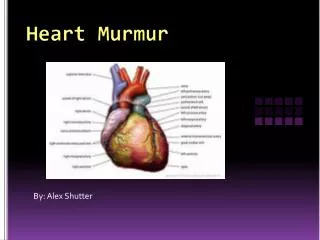
MURMUR AND Dynamic Auscultation OF Cardiovascular System. ANKUR KAMRA. Defining a heart murmur. A cardiac murmur is defined as a relatively prolonged series of auditory vibrations of Varying intensity (loudness), frequency (pitch), quality, configuration, and duration.

E N D
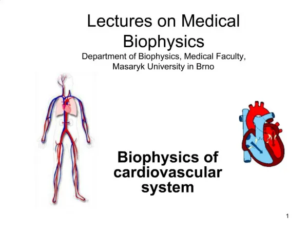
MURMUR AND Dynamic Auscultation OF Cardiovascular System ANKUR KAMRA
Defining a heart murmur A cardiac murmur is defined as a relatively prolonged series of auditory vibrations of Varying intensity(loudness), frequency (pitch), quality, configuration, and duration
How is a murmur produced? • Sound is produced by vibration • Vibration is generated by turbulence • Turbulence generated in the blood column set up vibrations in the vessel wall & cardiac structures causes murmurs
Leatham has attributed the production of murmurs or turbulence to three main factors: (1) high flow rate through normal or abnormal orifices, (2) forward flow through a constricted or irregular orifice or into a dilated vessel or chamber, and (3) backward or regurgitant flow through an incompetent valve, septal defect, or patent ductus arteriosus. Frequently, a combination of these factors is operative
CLASSIFICATION • Can be classified into organic, functional and innocent. • Organic refer to structural defect responsible for murmur • Important is that the term innocent and functional are not interchangeable. • Functional murmur should subserve a function like increased flow across aortic valve as in severe AR. • While innocent occur in absence of abnormalities of heart and circulation, more common in children and on right side.
Description of a Murmur • Position in the cardiac cycle • Site of murmur ] • Shape of murmur • Intensity • Quality & Pitch • Conduction • Dynamic changes
Dynamic Auscultation Listening to the change in character, behaviour and the intensity of the heart sounds and murmurs to physiological and pharmacological maneuvers……. “AUSCULTATE WITH ALTERED HEMODYNAMICS”
Conditions and interventions • Respiration • Postural Change • Valsalva maneuver • Exercise • Change in Cardiac Cycle length • Pharmacological agents.
What happen during respiration • Normal inspiration → • ↑ venous return to right side of the heart due to fall in intra thoracic pressure → ↑ stroke volume of right side • Dilatation of pulmonary vascular system causing decrease in pulmonary impedance there by increasing pulmonary hang out interval(>80 ms) • So leads to accentuation of R side murmur
RESPIRATION CONTINUE • Normal expiration → ↓ lung volume → ↑ pulmonary venous flow • Therefore, left sided murmurs are loudest during expiration except MR which remain unchanged. • While no change is seen • When complicated by RVF as due to high RVEDP no increase in venous return. • Aortic valvular ES do not vary with respiration • MR murmur do not vary with respiration
RESPIRATION CONTINUE • Assess changes during normal respiration • Patient should be in semiupright or sitting posture • In RV failure and PHT, no increase in venous return with inspiration, hence no inspiratory augmentation of right sided murmurs and gallops • Absence of respiratory influence is of no particular diagnostic value. • Effects of inspiration may be accentuated by Muller maneuver.
STANDING Auscultation is carried out immediately before and after the change in posture since effects may be quite transient persisting for only 10 – 15 heart beats If patient is unable to sit upright or stand, rapid application of tourniquets at upper thigh level may reduce venous return reproducing similar response
STANDING Rapid standing or sitting up from lying position or rapid standing from squatting posture results in • decreased venous return due to venous pooling in legs and splanchnic vessels leads to- • decreased stroke volume • decreased mean arterial pressure • decrease in heart size • followed by reflex increase in heart rate & systemic resistance
standing • All murmurs decrease except- • ESM of HOCM becomes louder and longer • Click occurs earlier, murmur becomes longer in MVP while loudness shows variable response
Sudden assumption of LYING DOWN POSITIONPASSIVE ELEVATION OF LEGS Increase in venous return → increase R.V. stroke volume → later after several cardic cycles left ventricle volume also increase . So Systolic murmur of AS,PS,MR, TR & VSD increase. MVP & HOCM murmurs decrease due to increase in LVEDV and LV size.
Squatting Sudden change from standing to squatting leads to- • Increases venous return & Stroke Volume • Increase of systemic vascular resistance due to kinking of iliac artery and reduction of pressure of gravity • Increase of systemic Arterial pressure with transient bradycardia
Squatting • Increased venous return and CO - augments most murmurs (AS,PS,MR,AR,VSD) Right heart murmurs do so earlier • Increased left ventricular volume - decreases murmur of HOCM and delayed murmur and click of MVP • Ejection murmur of TOF↑ due to increase pulmonary blood flow and decrease in right to left shunt
Other postural changes Assumption of L Lateral Position Causes closeness of heart to chest wall and transient rise of HR. So leads to – Increased murmur of MS, MR and austin flint murmur of AR Early appearance of click and systolic murmur of MVP due to increasec HR.
Sitting up and leaning forward causes more closeness of base of heart to chest wall so AR and PR murmurs more readly audible PRONE POSITION & KNEE CHEST POSITION Bring heart close to chest wall making pericardial rub more prominent
Valsalva Maneuver Relatively deep inspiration followed by forced exhalation against a closed glottis for 10 to 20 seconds Physician has to keep flat of the hand on the abdomen to provide the patient a force to breathe against
VALSALVA MANEUVER Forced expiration against closed glottis Manometer method: Patient blows into the mercury manometer and maintains 40 mmHg for 15 seconds Valsalva equivalent: Patient pushes back against examiner’s hand which is pressed downward on mid abdomen. The maneuver is demonstrated and patient practices the maneuver before assessment of murmur Caution : Not to be performed in IHD as it will reduce Coronary Blood Flow.
PHASES OF VALSALVA • PHASE 1-due to increase in intra thoracic pressure there is transient rise in LV output and systemic arterial pressure but there occurs fall in HR • phase 2(stain phase)- decrease in venous return first to right then to left leads to decrease in systolic, diastolic and pulse pressure and reflex tachycardia
PHASE 3- cessation of staining result in- • sudden increase in systemic venous return • but transient decrease in arterial pressure due to fall in intra-thoracic pressure • PHASE 4- return to pre valsalva • a transient overshoot of systemic arterial pressure • Reflex bradycardia
EFFECTS ON MURMUR • PHASE1 –as stroke volume fall there is decrease in – systolic murmur of AS, PS, MR, TR diastolic murmur of AR PR MS TS • PHASE2- reduction in LV volume and size leads to- increase in systolic murmur of HOCM increase in degree of MVP prolapse
PHASE 3- sudden increase in SVR leads to increase in right side murmurs • PHASE 4-left side murmur comes to control levels and may transiently increase. • ASD, MS and CHF – Phase 1 and 3 are normal but there is absence of decrease in arterial pressure tracing during phase 2 and overshoot of BP does not occur in phase 4 that leads to SQUARE WAVE RESPONSE ie. Instead of four phases there is only two phase.
The Muller Maneuver Converse of Valsalva Maneuver Less frequently employed Forcibly inspires while the nose is held closed and mouth is firmly sealed for about 10 sec. Augments murmur and filling sound originating in right side of the heart.
ISOMETRIC EXERCISE Use calibrated handgrip device or tennis ball or rolled up BP cuff. Measure the maximum effort. Patient exerts 70 – 100% of this maximum for about 30 seconds Simultaneous handgrip using both hands Valsalva maneuver during handgrip should be avoided
ISOMETRIC EXERCISE Hemodynamic changes: Significant increase in Arterial pressure Heart rate Cardiac output LV filling pressure LV size
Isometric Exercise • Systolic Murmur of AS reduced due to reduced gradient across aortic valve • AR , MR , VSD – increased due to increase systemic vascular resistance • MDM of MS – increased due to Increased CO • Syst Murmur of HOCM reduced • MVP murmur + click delayed
ISOMETRIC EXERCISE • Avoid in those with ventricular arrhythmias and myocardial ischemia • Contraindicated in recent myocardial infarction, uncontrolled hypertension, cerebrovascular disease, suspected aortic dissection
Cardiac cycle length changes post PVC and AF • ↑ preload will increases ventricular filling and size • Also in addition there is secondry increase in ventricular contractibilty of new beat and transient increase in arterial pressure.
Cardiac cycle length changes Increased ( L or R vent ejection murmurs ) AS PS HOCM(there is inc in SM but also decrese volume of pulse known as BROCKENBROUGH PHENOMENA) No change for MR , TR DM of AR increases due to transient rise in arterial pressure
Amylnitrite Inhalation Inhalation of Amyl Nitrate Crush ampoule in towel take 3-4 deep breaths over 10 – 15 secs Changes observed- • < 30 secs : Systemic vasodilatation • 30 – 60 secs : increase HR & CO • However majority of auscultaory changes are observed in first 30 sec
Due to increse in CO- SM of AS and PS SM of TR All functional SM DM of MS and TS DM of PR
Due to decrease in SVR following murmur are decreased- • SM of MR • DM and austin flint murmur of AR • SM of TOF • Due to decrease LV volume and size – • SM of HCM increases • early appearance of MVP click and murmur but softening of murmur occur due to decrease resistance to LV resistance
Amyl Nitrite Inhalation Augments Diminishes • Aortic stenosis Mitral regurgitation • Pulmonary stenosis TOF • Tricuspid regurgitation Mitral regurgitation • Mitral stenosis Austin Flint • Pulmonary regurgitation Aortic Regurgitation
Phenylephrine • ↑ BP & SVR ↓ CO & HR – last for 3-5mts • Reduces intensity of S1, A2-OS may widen • Augments the murmurs of VSD, PDA, MR, AR, TOF, Systemic AVF • Diminishes AS, MS & functional murmurs • ESM of HOCM diminishes • Click & murmur of MVP get delayed
Methoxamine & Phenylephrine Opposite effect of Amyl Nitrate Phenylephrine - due to short duration of action Systolic pressure elevated by 30 mm Hg for 3 to 5 mts EFFECT • Increases systemic arterial pressure • Reflex Bradycardia , decrease CO, decrease Contractility Caution : Not to be used in patients with CHF or Systemic hypertension.
Methoxamine & Phenylephrine AR , MR , VSD , TOF – Louder SM OF AS, PS and DM of PR and TS -show no changes LV size increases HOCM – Softer Click and Murmur of MVP - Delayed
Some general points about murmur before discussing individual murmurs
Sometime it is difficult to identify the timing of murmur • Murmur can be timed by simultaneous palpation of the carotid arterial pulse or by identifying S2 at base • Inching technique of Harvey and Levine • In tachycardia carotid sinus massage can slow down heart rate • In case of extra systole indentify the beat that follows pause and then first sound after pause will be S1
LENGTH • It generally reflect the pressure difference b/w two sites and this is true for all stenotic lesions like MS, AS, PS or TS • In regurgitant lesion length has no correlation with severity. • In AR length of murmur correlates better then MR but still not as reliable as stenotic lesions
CHARACTER • High frequency murmur occur when pressure difference b/w two chambers are high and low pressure difference has low frequency and pitch. • As a general rule regurgitant lesions are high frequency and stenotic are rough or low frequency. Murmur of AV stenosis are of low frequency while semilunar are of mixed frequency • High frequency or soft component of murmur is more widely audible this is reason why AS soft component is audible at apex and mistaken for MR • While low frequency or rough component is audible at site of best audibility of murmur.
Early Systolic murmurs Early systolic murmurs begin with S1 and extend for a variable period of time, ending well before S2 1. Acute severe mitral regurgitation • Regurgitation occurs into a normal-sized, relatively noncompliant left atrium and as LV-LA pressure gradient is abolished during late systole, termination of retrograde flow occurs well before S2. • best heard at apical impulse • Caused by: i. Papillary muscle rupture due to ishemia ii. Infective endocarditis -destruction of leaflet tissue, chordal rupture, or both