Download
1 / 130
1.3k likes | 1.46k Views
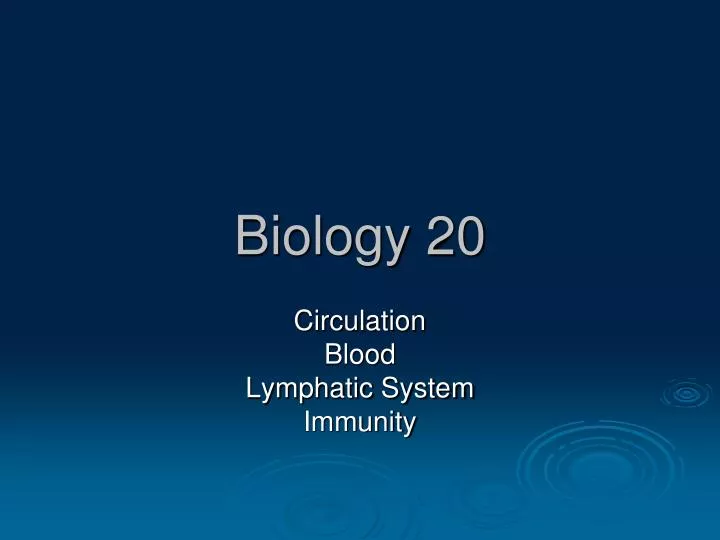
Biology 20. Circulation Blood Lymphatic System Immunity. The Importance of the Circulatory System. 96 000 km of blood vessels 60 trillion cells in body no cell is more than 2 cells away from a blood vessel. Functions of the Circulatory System. 3 main functions:

E N D
Biology 20 Circulation Blood Lymphatic System Immunity
The Importance of the Circulatory System • 96 000 km of blood vessels • 60 trillion cells in body • no cell is more than 2 cells away from a blood vessel
Functions of the Circulatory System • 3 main functions: 1) transports gasses, food, wastes, and hormones 2) carries molecules and cells that help defend against invading organisms 3) distributes heat throughout the body
Blood Vessels • Arteries • carry blood away from heart • elastic • stretch to accommodate blood surge from heart • pulse • thick walls to allow for stretch • high blood pressure
Blood Vessels • Arterioles • small arteries • arteries branch into many arterioles • connects to capillary • contains smooth muscle which can contract or relax according to nervous impulses • sympathetic nerve impulses affect diameter of arterioles: • Vasodilatation -- open • Vasoconstriction -- constricts
Blood Vessels • Capillaries • links arterioles to venules • site of exchange of nutrients and wastes between blood and interstitial fluid • I.F. is the fluid that surrounds and bathes body cells • smallest blood vessels • single layer of cells • big enough for only 1 RBC to pass through at a time • easily crushed = bruise
Blood Vessels • Venules • small veins • smooth muscle • large end of capillaries
Blood Vessels • Veins • carry blood towards the heart • larger diameter than arteries • lower pressure than arteries • (2 mmHg compared to 100 mmHg in arteries) • pull of gravity PROBLEM!! What forces the blood through a vein? • blood pressure is not high enough to “suck” blood back to the heart
Blood Vessels • Solution!! • Veins have one-way valves • only allow blood to move in one direction • skeletal muscles help veins in getting blood to the heart -- squeeze veins • wall is not as thick as arteries because blood pressure is lower – no surge of blood flow
Major Vessels of the Body • Superior Vena Cava • vein to heart from upper body • Inferior Vena Cava • Vein to heart from lower body • Pulmonary Artery • Artery from right ventricle to lungs • Pulmonary Veins (4) • Veins from lungs to left atrium • Aorta • Artery from left ventricle to body
The Heart • Structure • roughly in center of chest in thoracic cavity • protective membrane • pericardium forms a fluid filled sac to reduce friction • size of fist • two separate pumps • right and left • sides are separated by a septum which prevents mixing of oxygenated and deoxygenated blood
The Heart • Label the diagram of the heart. • Identify the following structures (next slide) • Outline the direction of flow of blood through the heart • Differentiate between oxygenated blood and deoxygenated blood by using red and blue arrows on diagram • List the general function of each structure.
Right Atrium Superior Vena Cava Inferior Vena Cava Right AV Valve (Tricuspid) Right Ventricle Pulmonary Artery Pulmonary Valve Pulmonary Vein Left Atrium Left AV Valve (Bicuspid) Left Ventricle Aorta Aortic Valve Coronary Arteries Interventricular Septum Chordae Tendinae The Heart
Superior Vena Cava Aorta Pulmonary Artery Pulmonary Veins Pulmonary Artery Left Atrium Pulmonary Veins Left AV Valve Right Atrium Left Ventricle Inferior Vena Cava Right Ventricle Pulmonary Valve Aortic Valve Right AV Valve Septum Semilunar Valve Semilunar Valve
The Heart’s Structures • Atria • Structure • thin walled chamber • Function • right atria - collect deoxygenated blood from body and head, and pass to ventricles. • left atria - oxygenated blood from the lungs, and pass it to ventricles.
The Heart’s Structures • Ventricles • Structure • thick muscular walls • larger than atria • one side is thicker than the other • Function • right ventricle pumps deoxygenated blood to lungs • left ventricle pumps oxygenated blood to body The left side has thicker walls than the right side
The Heart’s Structures • Interventricular septum • Wall of muscle that separates the right atrium and ventricle from the left atrium and ventricle.
The Heart’s Structures • Superior Vena Cava • Vein to right ventricle of the heart from upper body • Carries deoxygenated blood • Inferior Vena Cava • Vein to right ventricle of the heart from lower body • Carries deoxygenated blood • Aorta • Artery through which the left ventricle pumps blood to the body • Carries oxygenated blood
The Heart’s Structures • Pulmonary Artery • Pulmonary trunk – split into left and right arteries • Artery from right ventricle carries deoxygenated blood to lungs • Right pulmonary artery goes to right lung • Left pulmonary artery goes to the left lung. • Pulmonary Veins (4) • Veins from lungs carry oxygenated blood to left atrium • Right pulmonary vein from right lung • Left pulmonary vein from left lung
The Heart’s Structures • Atrioventricular valves (AV Valves) • The right AV valve is called the tricuspid valve and the left AV valve is called the mitral valve • Structure • flaps of tissue between atria and ventricles • attached to heart muscle by chords • chordae tendinae • cords prevent flaps from inverting • Function • Separate atria from ventricles • prevents back flow of blood from ventricles into atria
The Heart’s Structures • Pulmonary valve • Allows deoxygenated blood from right ventricle into pulmonary artery, but does not allow it to flow back into the ventricle. • Aortic valve • Allows oxygenated blood to flow from the left ventricle to the aorta, but not to return back to the ventricle. • Structure • Thin flap, half-moon shaped
AV Valves Open Atria Contract Ventricles Contract AV Valves Close The Heart’s Structures
Three Circulatory Systems 1) Pulmonary Circulation • From the heart to the lungs and back to the heart 2) Coronary Circulation • From the heart chambers to the heart muscles and back to the heart chambers • system of arteries, capillaries and veins within heart 3) Systemic Circulation • From heart to all other parts of the body and back to the heart.
Systemic Circulation • Involves 2 smaller systems • Renal Circulation • To the kidneys • Hepatic Circulation • From the digestive tract, through liver, then to heart • Important vessels • Carotid Arteries and Jugular Veins • head • Brachial Artery and Brachial Vein • arms • Femoral artery and Femoral Vein • legs
Biology 20 Heart Beat Heart Sounds Heart Rate Blood Pressure
Initiating the Heart Beat • Cardiac muscle contracts without external nervous stimulation • Sino-Atrial Node (SA Node) • Pacemaker • Coordinates contractions • Special muscle and nerves located in the right atrium • 70 beats/minute
Initiating the Heart Beat • The SA node sends nerve impulses: • across atria • contract now!! • To second node • AV node • Atrial-ventricular node
S-A Node A-V Node
Initiating the Heart Beat • AV node • mass of nerve tissue in septum • passes nerve impulse through septum to the bottom of ventricles • Impulse conducted upwards through nerves in the ventricles • Contraction of ventricles begins • Contraction begins at apex • Bottom to top contraction • Result… • Atria contract followed by the ventricles.
Setting the Heart’s Rate • Autonomic Nervous System in charge! • Sympathetic nerves • stimulated during times of stress • Increases heart rate • Parasympathetic nerves • stimulated during times of non-stress • returns heart to slow rate
Setting the Heart’s Rate • Problem: • SA node malfunction • Ventricular fibrillation • disorganized and random contraction of heart cells • no longer pumps blood • death • Solution: • First -- Electro shock to halt the “short circuit” • Then -- installation of an artificial pacemaker
Electrocardiogram • Electrodes are placed on the body surface • Connected to a recording device • Electrical impulses from heart are displayed on a screen. • P wave • atrial contraction • QRS wave • ventricular contraction • Used to diagnose heart problems • more evident during heavy exercise • Doctors identify dead patches of heart muscle which will not conduct impulses.
Cardiac Cycle • One heart beat • Systole • ventricles contract • Diastole • ventricles relax • 70 - 75 beats/min • two sides of heart beat in unison • first the atria then the ventricles