Download
1 / 39
410 likes | 730 Views
Assessment of Cardiovascular System. NUR123 Spring 2009 K. Burger, MSEd, MSN, RN, CNE PPP by: Victoria Siegel, RN, CNS, MSN & Sharon Niggemeier, RN, MSN Revised by: Kathleen Burger. Cardiovascular Anatomy & Physiology. Heart is shaped like “Cone” “top” of the heart is the base

E N D
Assessment of CardiovascularSystem NUR123 Spring 2009 K. Burger, MSEd, MSN, RN, CNE PPP by: Victoria Siegel, RN, CNS, MSN & Sharon Niggemeier, RN, MSN Revised by: Kathleen Burger
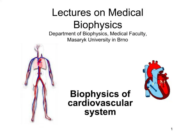
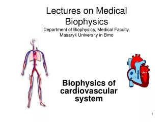
Cardiovascular Anatomy & Physiology • Heart is shaped like “Cone” • “top” of the heart is the base • “bottom” is the apex • Heart size = clenched fist • Precordium- area on anterior chest that covers heart and great vessels • Atria are tilted slightly toward the back and ventricles extend to left and toward anterior chest wall.
Unoxygenated Blood: Superior Vena Cava& Inferior Vena Cava R Atrium Tricuspid valve R Ventricle Pulmonic Valve Pulmonary Artery to lungs (gets oxygenated) Oxygenated Blood: Pulmonary veins L Atrium Mitral Valve L Ventricle Aortic Valve Aorta Body Cardiovascular: Blood Flow
Cardiovascular: Blood SUPPLY • There are two main coronary arteries, the left (LCA) and the right (RCA) • Coronary artery blood flow to the myocardium occurs primarily during diastole, when coronary vascular resistance is minimized. • To maintain adequate blood flow through the coronary arteries, the diastolic pressure must be at least 60 mmHg.
Cardiovascular: Cardiac Cycle • 2 phases • DIASTOLE: AV valves open – passive flow (75% of volume) into relaxed ventricles, then atria contract – active flow of remaining 25% into ventricles • SYSTOLE : AV valves close, ventricle pressure increases, ventricle contracts, Seminular valves open, blood pumped into pulmonary and systemic arteries
Cardiovascular: Heart Sounds • Heart sounds: lub dub • SYSTOLE: lub= S1 (closing of AV valves) • DIASTOLE: dub = S2 (closing of semilunar valves) • During the cardiac cycle, valves are opening and closing, causing different heart sounds (S1 and S2). • Sometimes abnormal heart sounds are heard due to improper opening or closing of the valves.(murmurs)
Cardiovascular: Heart Sounds • Characteristics of Heart Sounds • Frequency (pitch): high or low • Intensity (loudness): loud or soft • Duration: very short heart sounds or longer periods of silence • Timing: systole or diastole
Cardiovascular: Conduction • Heart contracts by itself through its own conduction system: • Sinoatrial (SA)node – (pacemaker) initiates electrical impulse • AV node • Bundle of HIS (L & R Bundle Bbranches) • Purkinje fibers
Cardiovascular: Conduction • Electrical impulses shown on ECG (EKG) • PQRST wave correlates to impulses traveling through the heart. • SA to AV = P wave, (atrial stimulation) • Stimulus spreads through bundle of His = QRS complex • Repolarization of ventricles =T wave on
Cardiovascular:Pumping Ability • Cardiac Output (C.O.) = volume of blood in liters ejected by the heart each minute. • Adult = 4-6 liters/minute • CO = HR x SV • Heart Rate (HR) = number of times ventricles contract each minute. • Stroke Volume (SV) = The amount of blood ejected by the left ventricle during each systole.
Cardiovascular • Preload = degree of stretch of myocardial fibers at end of DIASTOLE. The more the heart is filled (within limits, i.e., not over-filled), the more forcefully it contracts. • Afterload = pressure or resistance the ventricles must overcome to pump out blood. The amount of resistance is directly related to arterial blood pressure and the diameter of the vessels.
Assessment: Subjective • Personal and family history • Diet history: 24 hr. sample diet Opportunity for teaching food selection and preparation • Socioeconomic status – ability to purchase proper foods, medicines. Employment and its effects on health? • Cigarette smoking : # packs /day and also # years smoked PACK YEARS
Assessment: Subjective • Physical Activity/Inactivity – 30 minutes daily of moderate exercise recommended on most days ( Healthy People 2010 ) • Obesity – associated with HTN, hyperlipidemia, and diabetes and all contribute to CV disease. • Type A personality – not conclusive proof • Current Health Problems – describe health concerns.
Assessment: Subjective • Chest pain: or discomfort, a symptom of cardiac disease, can result from ischemic heart disease, pericarditis and aortic dissection. • Chest pain: can also be due to non- cardiac causes; pleurisy, pulmonary embolus, hiatal hernia and anxiety musculoskeletal strain, GERD
Assessment- Chest Pain • Onset • Duration • Frequency • Precipitating factors / Relieving factors • Location • Radiation • Quality • Intensity
Assessment: Subjective • Paroxysmal Nocturnal Dyspnea – client has been recumbent for several hours, increase in venous return leads to pulmonary congestion. • Fatigue- resulting from decreased cardiac output is usually worse in evening. Ask pt. if can they perform same activities as a year ago
Assessment: Subjective • Palpitations- fluttering or unpleasant awareness of heartbeat. Non- cardiac- causes- fatigue, caffeine, nicotine, alcohol • Weight gain- a sudden increase in wt. of 2.2 pounds (1 kg) can be result of accumulation of fluid (1L) in interstitial spaces, known as edema. • Syncope- transient loss of consciousness, decrease in perfusion to brain.
Assessment:ObjectiveBeginning Inspection • General appearance: Build, skin color, LOC, presence of SOB, DOE • Older age? • Transcultural considerations? • Skin- color and temperature – look for symmetry in color, temp, any cyanosis? • Extremities – assess skin changes, vascular changes, clubbing, capillary filling and edema. • Neck vein distention?
Assessment:Objective • BP: supine – change position 1-2 minutes, check again. • Normally, systolic drops slightly or remains unchanged and diastolic increases slightly. • Carotid &Peripheral pulses are assessed for: • Presence • Amplitude • Rhythm • Rate • Equality
Assessment:Objective • Specific assessments for particular populations: • Assessment for Infants • Assessment for Children • Assessment for Pregnant Females • Assessment for Elderly,
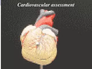
Assessment:Objective • Precordium Assessment- area over heart, done by: • Inspection • Palpation • Percussion • Auscultation
Physical Assessment • Inspection- side to side, at right angle and downward over precordium where vibrations are visible. • Point of Maximal Impulse (PMI) Apical Impulse – located at 5th intercostal (IC) space at midclavicular line (MCL) – mitral area • Right Ventricular (RV area) • Epigastric area • Pulmonic area
Physical Assessment • Palpation: fingers and most sensitive part of palm of hand to detect any precordial motion or thrills. • Palpate apical impulse • Percussion: estimate heart size, most accurately done by chest x-ray • Auscultation:– evaluates heart rate, rhythm, cardiac cycle and valvular function.
Physical Assessment: Auscultation • Diaphragm of stethoscope – 1st and 2ndheart sounds and high frequency murmurs. lub-dub • Use bell of stethoscope – low frequency gallops and murmurs. • Paradoxical splitting of S2 – severe myocardial depression, may be seen with an MI, aortic stenosis or other causes.
Auscultation:EXTRA Heart Sounds • S3 (Ventricular Gallop): rapid, passive filling phase during diastole into ventricle. • S4 (Atrial Gallop): active filling of ventricles with “atrial kick”. Pathologic, may be heard with advancing age because of stiffened ventricle. • Both S3 and S4 = Summation Gallop: indication of severe heart failure. • Murmurs – Turbulent blood flow through normal or abnormal valves.
AuscultationEXTRA HEART SOUNDS • Murmurs – are classified according to their timing: Systolic or diastolic and loudness: grading • Innocent systolic between S1 and S2 commonly heard in children and adults under 30. • Configuration ( description )of murmurs:Pitch, quality, location, radiation, posture, quality (Crescendo- Decrescendo)
Intensity of murmur: Grade 1: faint 2: soft 3: moderately loud 4: loud with thrill 5: very loud (stethoscope partially off chest) 6: stethoscope off chest, thrill AuscultationEXTRA HEART SOUNDS
AuscultationEXTRA HEART SOUNDS • Pericardial Friction Rubs- results from inflammation of pericardial membrane. • Ejection Click- Early systole, stiff deformed valve, high pitch, apex, diaphragm. • Opening snap – Immediately after S2 stenotic mitral or tricuspid valve leaflets recoil abruptly during diastole.
CARDIAC AUSCULTATIONSTEPS • Palpate PMI (5th ICS, midclavicular) and place your stethoscope = MITRAL area • Count rate, assess rhythm • Isolate S1 ( use carotid pulse prn )& listen. S1 should be louder than S2 in this area • Move stethoscope to left sternal border (LSB) in the same ICS = TRICUSPID area. Listen. S1 should still be louder than S2 in this area • Move stethoscope up LSB to 3rd ICS = ERBs POINT. S1 =S2 in this area
CARDIAC AUSCULTATIONSTEPS (continued) • Continue up LSB to 2nd ICS to PULMONIC area. S2 should be louder than S1 in this area • Move stethoscope to RSB 2nd ICS to listen at the AORTIC area. S2 is louder than S1 • Listen at MITRAL and TRICUSPID areas for S3 and S4. Use the BELL of stethoscope. • Repeat steps 1-7 listening exclusively for murmurs in all 5 areas. Use DIAPHRAGM AND BELL. • Do auscultation steps supine, lateral,& sitting
Assessment : Subjective • Leg Pain: • Hx: DVT? • Arm/leg skin changes,varicose veins • Edema • Medications
Assessment : Objective • Inspection: • skin including color & hair distribution • skin ulcers? • symmetry in extremity size? • jugular vein distention • varicosities? • Palpation: • pulses, tenderness, temperature, edema, capillary refill
Assessment:Objective • Pulses- carotid, brachial, radial, femoral, popliteal, posterior tibialis and dorsalis pedis. • 0= nonpalpable • 1+ = easily obliterated • 2+ = weak, but cannot be obliterated • 3+ = easy to palpate; full; cannot be obliterated. • 4+ = strong, bounding; may be abnormal
Assessment : Objective • Edema- Check for pretibial edema. How high up does it go? • 1+- Mild pitting, slight indentation. • 2+- Moderate pitting- indentation subsides rapidly. • 3+- Deep pitting, indentation remains short time, leg looks swollen. • 4+- Very deep pitting, very swollen.
Assessment : Objective • Allen test- occlude radial & ulnar arteries, pt. opens and closed fist, hand should blanch. Then let go of ulnar arteryquickly while you are occluding radial artery; if hand turns pink, ulnar is intact.
Figure 5-37. Auscultation: Carotid arteries in older adults; Use bell of stethoscope
Doppler Assessment • Position client supine • Externally rotate leg • Apply conducting gel • Place transducer over pulse site • 45 degree angle with light pressure • Listen for whooshing sound
Summary: Cardiovascular • Physical assessment Includes: • Neck vessels • Precordium • Inspection and palpation of peripheral system with auscultation of the carotids