Download
1 / 23
340 likes | 909 Views
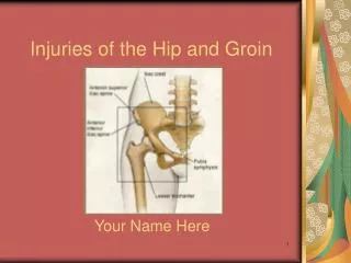
Injuries Of The Hip And Femur. Dislocations Of The Hip. Complete loss of contact between the articular surfaces forming the joint. Classified according to the direction of the femoral head to: Posterior ( commonest ) . Anterior. Central (comminuted or displaced fracture of acetabulum ).

E N D
Dislocations Of The Hip • Complete loss of contact between the articular surfaces forming the joint. Classified according to the direction of the femoral head to: • Posterior (commonest). • Anterior.
Posterior Dislocation • Four out of five traumatic dislocations. • Mechanism of injury: Road traffic accident when the victim thrown forward striking the knee against the dashboard with the hip and knee flexed forcing the head out of the acetabulum, some time posterior wall fracture happen (fracture dislocation).
Clinical Features • The leg is shortened, adducted, internally rotated and flexed at the hip joint. • Be careful if associated with fracture femur (rule). • Always examine for signs of sciatic nerve injury.
Radiological evaluation • X-ray: • AP view of the pelvis is a must in all severely traumatized patient. • The head is out and above the acetabulum. • Search for acetabular rim or femoral head fracture. • CT scan: show the anatomy of concomitant fractures, exclude intra articular bone piece.
Thompson and epstein classification • Type I: dislocation with no or small chip fracture. • Type II: with large posterior wall fracture. • Type III: with posterior wall cominution. • Type IV: with fracture acetabular floor. • Type V: with fracture femoral head.
Treatment • Urgent reduction under GA (assistant stabilize the pelvis, traction applied then flexion of hip and knee to 90◦then gradual rotation till you here clunk of reduction. • X-ray is essential to confirm reduction and exclude fractures.
After Treatment • Type I: Traction for 3 weeks then partial wt. bearing. • Type II: Open reduction and rigid fixation of posterior wall followed by traction for 6 weeks. • Type III: Traction for 6 weeks. • Type IV&V: closed reduction may lead to automatic reduction of the fractures, if not open reduction and internal fixation followed by traction for 6 weeks. • Full weight bearing allowed only after 12 weeks.
Complications Early: • Sciatic nerve injury. • Vascular injury (superior gluteal artery). • Associated fracture of femoral shaft. Late: • Avscular necroses: Immediate reduction 10%. Delayed reduction 40%. • Myosits ossificans. • Unreduced dislocation. • Osteoarthrits.
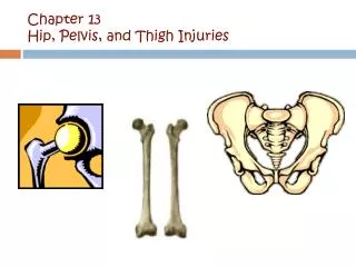
Hip Fractures • Involves head, neck and subtrochanteric fractures. • Femoral neck fractures involve the narrow neck between the round head of the femur and the shaft. • Neck fractures are classified into (vascularity, treatment, complications): • Intra capsular. • Extra capsular.
Intra Capsular Fractures • Fracture happen in the intracapsular part of the neck. • Common in the seventh and eighth decade. • Risk factors: • Osteoporoses and osteomalacia. • Diabetes mellitus. • Alcoholism. • Chronic debilitating diseases.
Mechanisms of injury • Fall on the greater trochanter directly. • Catching the toe in the carpet and twisting the hip to external rotation. • In young patients (FFH,RTA).
Clinical features • Fall followed by pain in the hip. • The patient lies with the limb externally rotated and short. • Young patients usually have associated severe injuries of the lower limb( fracture femur).
X-ray: This is divided according to the level of the fracture line in the neck as follows: 1) Sub capital. 2) Trans cervical. 3) Basal. • Stress fractures and impacted fractures may show no finding.
Fracture displacement is assessed by the garden classification: • Garden I : incomplete fracture of the neck (so-called abducted or impacted). • Garden II : complete without displacement.
Garden III: complete with partial displacement; fragments are still connected by posterior retinacular attachment; there is malalignment of the femoral trabeculae( adducted). • Garden IV : this is a complete femoral neck fracture with full displacement; the proximal fragment is free and lies correctly in the acetabulum so that the trabeculae appear normally aligned.
Treatment Initial traction to relief pain till the definitive treatment. Fractures at this level have a poor capacity for union due to the following factors: A) Interference with the blood supply to the proximal fragment. B) Difficulty in controlling the small proximal fragment. C) The lack of organization of the fracture hematoma due to the presence of the synovial fluid. D) Lack of periostium. • Because of these facts there is no role of conservative treatment.
Surgical Treatment • All patients under the age of 70 year by: Closed Reduction And Internal Fixation. Two essential principles to be followed in the surgical management of this fracture are: (A) Perfect anatomical reduction. (B) Rigid internal fixation. • The principle is to do closed reduction under image intensifier and then internal fixation either by:
Multiple compression canulated screws. • Dynamic Hip Screw.
Patients above 70year: • Partial hip replacement (bipolar,Austin-moor). • Total hip replacement.
Complications • General (DVT, PE, pneumonia, bed sores). • Avascular necroses of the femoral head. • Non union. • Osteoarthritis.