Download
1 / 1
10 likes | 231 Views
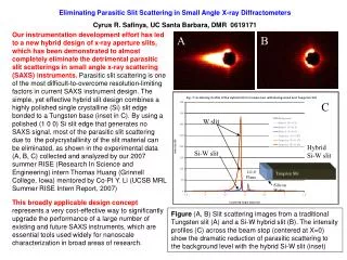
L. 2r. 200 nm. 5.00 μ m. Fig 6: Two pore structures that yield similar PSDs, but have vastly different SAXS curves. Small Angle X-Ray Scattering from Nanoporous Biocarbon Mikael Wood 1 , Jacob Burress 1 , Peter Pfeifer 1 , Jan Ilavsky 2

E N D
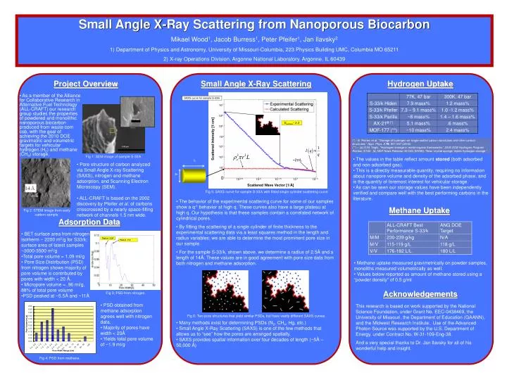
L 2r 200 nm 5.00 μm Fig 6: Two pore structures that yield similar PSDs, but have vastly different SAXS curves • Small Angle X-Ray Scattering from Nanoporous Biocarbon • Mikael Wood1, Jacob Burress1, Peter Pfeifer1, Jan Ilavsky2 • Department of Physics and Astronomy, University of Missouri-Columbia, 223 Physics Building UMC, Columbia MO 65211 • X-ray Operations Division, Argonne National Laboratory, Argonne, IL 60439 Project Overview Small Angle X-Ray Scattering Hydrogen Uptake • As a member of the Alliance for Collaborative Research in Alternative Fuel Technology (ALL-CRAFT) our research group studies the properties of powdered and monolithic nanoporous biocarbon produced from waste corn cob, with the goal of achieving the 2010 DOE gravimetric and volumetric targets for vehicular hydrogen (H2) and methane (CH4) storage. SAXS curve for sample S-33/k 107 106 Dsurface~ 2.3 105 104 Scattered Intensity [1/cm] (*) - E. Poirier, et al, “Storage of hydrogen on single-walled carbon nanotubes and other carbon structures.” Appl. Phys. A 78, 961-967 (2004). (**) – (a) O.M. Yaghi, “Hydrogen storage in metal-organic frameworks.” 2006 DOE Hydrogen Program Review, ST22. (b) NSF News Release 06-043 (3/9/06): “New ‘crystal sponge’ triples hydrogen storage.” 103 Fig 1: SEM image of sample S-33/k 102 • The values in the table reflect amount stored (both adsorbed and non-adsorbed gas). • This is a directly measurable quantity, requiring no information about nanopore volume and density of the adsorbed phase, and is the quantity of foremost interest for vehicular storage. • As can be seen our storage values have been independently verified and compare well with the best performing carbons in the literature. ~2π/L • Pore structure of carbon analyzed via Small Angle X-ray Scattering (SAXS), nitrogen and methane adsorption, and Scanning Electron Microscopy (SEM). • ALL-CRAFT is based on the 2002 discovery by Pfeifer et al. of carbons crisscrossed by a nearly space-filling network of channels 1.5 nm wide. 101 10-2.5 10-2 10-1.5 10-1 10-.5 Scattered Wave Vector [1/Å] Fig 5: SAXS curve for sample S-33/k with fitted single cylinder scattering curve • The behavior of the experimental scattering curve for some of our samples show a q-1 behavior at high q. These curves also have a large plateau at high q. Our hypothesis is that these samples contain a correlated network of cylindrical pores. • By fitting the scattering of a single cylinder of finite thickness to the experimental scattering data via a least squares method in the length and radius variables; we are able to determine the most prominent pore size in our sample. • For the sample S-33/k, shown above, we determine a radius of 2.5Å and a length of 14Å. These values are in good agreement with pore size data from both nitrogen and methane adsorption. Methane Uptake Fig 2: STEM image from early carbon sample. Adsorption Data • BET surface area from nitrogen isotherm ~ 2200 m2/g for S33/k; surface area of latest samples ~3000-3500 m2/g • Total pore volume = 1.09 ml/g • Pore Size Distribution (PSD) from nitrogen shows majority of pore volume is contributed by pores with width < 20 Å. • Micropore volume = .96 ml/g, 88% of total pore volume • PSD peaked at ~5.5Å and ~11Å Peak at ~5.5Å Peak at ~11Å • Methane uptake measured gravimetrically on powder samples, monoliths measured volumetrically as well. • Values below reported as amount of methane stored using a “powder density” of 0.5 g/ml Acknowledgements Fig 3: PSD from nitrogen. • PSD obtained from methane adsorption agrees well with nitrogen data. • Majority of pores have width < 20Å • Yields total pore volume of ~1.9 ml/g This research is based on work supported by the National Science Foundation, under Grant No. EEC-0438469, the University of Missouri, the Department of Education (GAANN), and the Midwest Research Institute. Use of the Advanced Photon Source was supported by the U.S. Department of Energy, under Contract No. W-31-109-Eng-38. And a very special thanks to Dr. Jan Ilavsky for all of his wonderful help and insight. • Many methods exist for determining PSDs (N2, CH4, Hg, etc.) • Small Angle X-Ray Scattering (SAXS) is one of the few methods that allows us to “see” how the pores are arranged spatially. • SAXS provides spatial information over four decades of length (~5Å – 50,000 Å) Fig 4: PSD from methane.