Download
1 / 40
410 likes | 672 Views
Cells and organs of the immune system. IL-3. GM-CSF, IL-3. p. 25. IL-7, IL-2, IL-4. various growth factors and cytokines. IL-2, IL-4 IL-5, IL-6. Cell-culture systems can support the growth and differentiation of lymphoid cells. p. 26. Hematopoietic growth factors

E N D
IL-3 GM-CSF, IL-3 p. 25 IL-7, IL-2, IL-4 various growth factors and cytokines IL-2, IL-4 IL-5, IL-6
Cell-culture systems can support the growth and differentiation of lymphoid cells p. 26
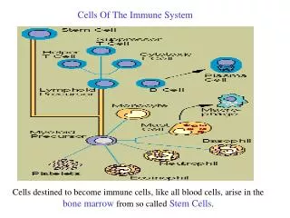
Hematopoietic growth factors Colony-stimulating factors support growth of specific cell types IL-3 (multilineage) EPO (erythropoietin)- RBCs Target cells must possess receptors for the appropriate cytokine Earliest cells are “pluripotent”: can develop into many types of cells CD34 is “marker” for this cell type (p. 31)
Regulation of hematopoiesis Production of cytokines by bone-marrow stromal cells Production of cytokines by other cells Expression of receptors Programmed cell death (apoptosis) important general mechanism for homeostasis
Immune cells have different life spans Neutrophils- a few days Memory cells-years Apoptosis is important for regulation of cell numbers
p53 defects implicated in many cancers (tumor suppressor gene) p. 30 Overproduction of bcl-2 implicated in B-cell lymphoma
Distribution of blood cells in healthy adult p. 32 p. 35
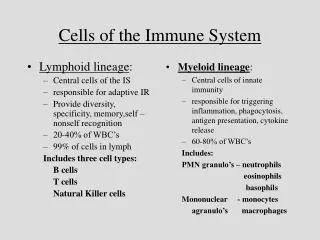
Cells of the immune system Lymphoid cells B cells T cells “null cells” (no antigen receptors) primarily NK cells
B lymphocytes Express membrane bound immunoglobulin (Ig; antibody) approx. 150000 molecules per cell all have the same specificity When activated by antigen these become plasma cells secrete immunoglobulin live 1-2 weeks some become memory cells
T lymphocytes Also have membrane-bound, antigen-specific receptor Antigen-binding part is organized similarly to B cell antigen receptor (the rest is different) B cell can recognize free antigen T cell cannot
T cell antigen recognition Antigen must be processed and “presented” to T cell by an antigen-presenting cell (APC) Antigen must be bound to MHC molecule on the APC T cell must directly contact the APC (antibodies can circulate) Memory T cells are produced
T cell subsets I. Helper T cells (TH cells) Express CD4 recognize antigen bound to MHC Class II These cells release cytokines which stimulate their proliferation and effector functions of other cells TH1 and TH2 subsets control different parts of the immune response
II. Cytotoxic T cells express CD8 are MHC-Class I restricted respond to “altered-self” cells i.e., tumor cells and virus-infected cells don’t secrete many cytokines release toxic granules that kill target cells
Null cells no antigen-specific receptors no specificity Natural killer (NK) cells are most prominent- about 5-10% of all lymphocytes Thought to deal with cells that have reduced levels of MHC Class I Kill target cells with release of toxic granules “First line” of defense “NK1-T cell”: seen experimentally when establishing T cell lines
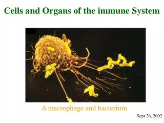
Mononuclear cells • Monocytes in blood • Macrophages in tissue (monocytes migrate • after a few hours in the blood) • Classical names based on where • macrophages reside • Kupffer cells in liver • Histiocytes in connective tissue • Alveolar macrophages in lung, etc.
Macrophages (an other phagocytes) are stimulated by: encountering antigen cytokines Stimulated macrophages enhance immune response better killers release more inflammatory molecules activate more T cells (express more MHC Class II
Some pathogens can resist these Mycobacterium tuberculosis Listeria monocytogenes Neisseria gonorrhoeae Candida albicans among others
Granulocytes (so named due to histology) Neutrophils short-lived early response; active in inflammation phagocytes Eosinophils also phagocytes much rarer and less important in inflammation thought to play a role in defense against parasites
Basophils not phagocytic release pharmacologically active substances from their granules prominent in allergic responses Mast cells differentiate in tissue, not bone marrow found in tissue such as skin, digestive, genitourinary tract, etc. release substances similar to those of basophils
Dendritic cells- have long processes that resemble dendrites Express MHC Class II and “co-stimulatory” cell surface molecules (such as B7) Very good at presenting antigen Are of hematopoietic lineage
Types and locations of dendritic cells Langerhans cells- skin Interstitial dendritic cells- in most organs Interdigitating- in thymus and other lymphoid organs Circulating
Primary and secondary lymphoid organs Primary- maturation of lymphocytes thymus bone marrow Secondary- trap antigen and allow interaction with lymphocytes lymph nodes spleen MALT (mucosal-associated lymphoid tissue GALT (gut etc.)
Lack of functional thymus Many fewer circulating T cells No cell-mediated immunity Increased susceptibility to viral and fungal infections Aging causes decline in thymus function
Secondary lymphoid tissue lymphoid follicles primary- resting B cells and APCs secondary- proliferating B cells Lymph nodes and spleen are most organized
Plasma cells In medulla (center) p. 48
Spleen Lymph nodes trap circulating antigen exchange between lymphatic vessels and blood Spleen filters antigens from blood Organization of spleen “red pulp”- old RBCs and macrophages “white pulp” marginal zone- B cells PALS surrounds artery, contains T cells (T cells are activated first)
MALT Tonsils Peyer’s patches Appendix “IELs” – intraepithelial lymphocytes T cells have unusual receptors M cells “deliver” antigen to immune cells
antigen p. 51
Skin (primary barrier, remember?) Also has some specialized cells Keratinocytes can produce cytokines can express MHC Class II Langerhans cells- APCs Lymphocytes seem to have limited repertoire many have “alternative” T cell receptor