Download
1 / 40
460 likes | 717 Views
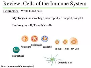
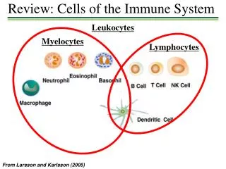
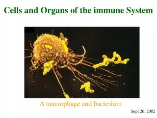
Chapter 2. Cells and Tissues of the Immune System. CELLS OF THE IMMUNE SYSTEM. Phagocytes. Phagocytes, including neutrophils and macrophages , are cells whose primary function is to identify , ingest , and destroy microbes In host defense consist of sequential steps:

E N D
Chapter 2 Cells and Tissues of theImmune System
Phagocytes • Phagocytes, including neutrophils and macrophages, are cells whose primary function is to identify, ingest, and destroy microbes • In host defense consist of sequential steps: • recruitment of the cells to the sites of infection, • recognition of and activation by microbes, • ingestion of the microbes by the process of phagocytosis, and • destruction of ingested microbes
Neutrophils • Polymorphonuclear leukocytes (PMN), nucleus with three to five connected lobules • most abundant population of circulating WBC • 12 to 15µm • The cytoplasm contains granules of two types, specific granules (enzyme such as lysozyme, collagenase, and elastase) that do not stain strongly with either basic or acidic dyes and azurophilic granules (lysosomes containing enzymes and other microbicidal substances, including defensins and cathelicidins) • Production of neutrophils is stimulated by granulocyte colony-stimulating factor (G-CSF) • An adult human produces more than 1 x 10¹¹ neutrophils per day • function for a few hours (6h) and then die
Mononuclear Phagocytes (MMN) • Play central roles in innate and adaptive immunity • Unlike neutrophils, macrophages are not terminally differentiated and can division at an inflammatory site • Incompletely differentiated and is called monocyte • Some develop abundant cytoplasm and are called epithelioid cells • Later stages of the innate immune response, 1 or 2 days after infection • Macrophages in different tissues, microglial cells (CNS), Kupffer cells (liver), alveolar macrophages (pulmoner); osteoclast(bone)
Several important functions in innate and adaptive immunity: • A major function of macrophages in host defense is to ingest and kill microbes • ingest dead host cells as part of the cleaning up process after infection or sterile tissue injury • Activated macrophages secrete proteins, called cytokines, • Macrophages serve as APCs • promote repair of damaged tissues by stimulating new blood vessel growth (angiogenesis) and synthesis of collagen-rich extracellular matrix (fibrosis) • Macrophages are activated to perform their functions by recognizing many different kinds of microbial molecules as well as host molecules produced in response to infections (Toll-like receptors)
Macrophages can acquire distinct functional capabilities, depending on the types of activating stimuli • Some of these cytokines activate macrophages to become efficient at killing microbes, called classical activation • Other cytokines activate macrophages to promote tissue remodeling and repair, called alternative activation • Macrophage-like cells (hemocytes) are phylogenetically the oldest mediators of innate immunity
Mast Cells • Mast cells are bone marrow–derived cells that are present in the skin and mucosal epithelium and contain abundant cytoplasmic granules filled with cytokines,histamine, and other mediators • Stem cell factor (also called c-Kit ligand) is a cytokine that is essential for mast cell development • Express plasma membrane receptors for IgE and IgG antibodies • Provide defense against helminths but are also responsible for symptoms of allergic diseases
Basophils • Basophils are blood granulocytes with many structural and functional similarities to mast cells • less than 1% of blood leukocytes • Normally not present in tissues, basophils may be recruited to some inflammatory sites • Importance in host defense and allergic reactions is uncertain
Eosinophils • Blood granulocytes express cytoplasmic granules containing enzymes that are harmful to the cell walls of parasites but can also damage host tissues • GM-CSF, IL-3, and IL-5 promote eosinophil maturation from myeloid precursors
Antigen-Presenting Cells (APC) • Specialized to capture microbial, display them to lymphocytes, and provide signals to stimulate the proliferation and differentiation of lymphocytes • Major type of APC that is involved in initiating T cell responses is the dendritic cell (DC) • Macrophages present antigens to T cells during CMI responses, and B lymphocytes function as APCs for helper T cells during humoral immune responses • A specialized cell type called the follicular dendritic cell (FDC) displays antigens to B lymphocytes during particular phases of humoral immune responses • APCs link responses of innate immune system to responses of the adaptive immune system,
Dendritic Cells • Play important roles in innate immunity to microbes and in antigen capture and the induction of T lymphocyte responses to protein antigens • Arise from bone marrow precursors, mostly of the monocyte lineage, and are found in many organs • Activated DC also express molecule called costimulators, which function in concert with antigen to stimulate T cells
Follicular Dendritic Cells (FDC) • Are present involve in specialized collections of activated B cells, called germinal centers • Are not derived from precursors in the bone marrow and are unrelated to the dendritic cells • FDCs trap antigen-antibodies complexes to or complement products for recognition by B lymphocytes • Important for the selection of activated B lymphocytes
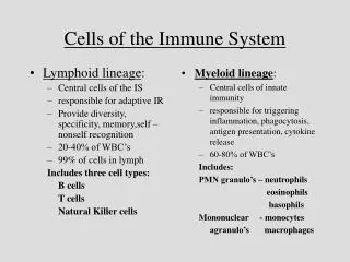
Lymohocytes • Lymphocytes are the only cells in the body capable of specifically recognizing and distinguishing different antigenic determinants and are responsible for the two defining characteristics of the adaptive immune response, specificity and memory • Major subsets of B cells (B2 cell) are follicular B cells, marginal zone B cells, and B1 cells, • T cell subsets are helper T lymphocytes (Th CD4+), cytotoxic T lymphocytes (CTLs), which express an antigen receptor called the αβ receptor, CD4+ regulatory T cells , and γδ T cells • NKT cells are a numerically small population of lymphocytes that share characteristic of both NK cells and T cells
Naive Lymphocytes • Are mature T or B cell emigrants from generative lymphoid organs that have never encountered foreign antigen • Die after 1 to 3 months, if they do not recognize antigens • Naive and memory lymphocytes, are both called resting lymphocytes, because they are not actively dividing, nor are performing effector functions
Effector Cells • Include helper T cells, CTLs, and antibody secreting B cells • Majority of differentiated effector T lymphocytes are short lived and not self- renewing • Many antibody-secreting B cells are morphologically identifiable as plasma cells
Memory Cells • Identified by their expression of surface proteins although it is still not clear which definitive markers of memory populations • Memory B lymphocytes express certain classes of membrane Ig, such as IgG, IgE, or IgA, whereas naive B cells express only IgM and IgD • In humans, CD27 expression is a good marker for memory B cells • In humans, most naive T cells express a 200-kD isoform of a surface molecule called CD45RA (for restricted A) • In contrast, most activated and memory T cells express a 180-kD isoform of CD45RO
Small lymphocyte Large lymphocyte
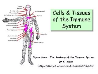
Lymphoid tissues are classified as generative organs or primary lymphoid organs (Thymus and BM), where lymphocytes first express antigen receptors and attain phenotypic and functional maturity, and as peripheral organs or secondary lymphoid organs (LN, spleen,…) where lymphocyte responses to foreign antigens are initiated and develop
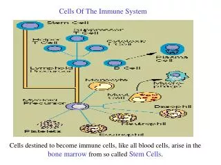
Bone Marrow • Is the site of generation of all circulating blood cells in the adult • During fetal development, the generation of all blood cells, called hematopoiesis • Stem cells express two proteins called CD34 and stem cell antigen 1(Sca-1) • Proliferation and maturation of precursor cells in the bone marrow are stimulated by cytokines are called colony-stimulating factors
Thymus • Is the site of T cell maturation • Is a bilobed organ situated in the anterior mediastinum. Each lobe is divided into multiple lobules by fibrous septa, and each lobule consists of an outer cortex and an inner medulla • Cortex contains a dense collection of T lymphocytes • Scattered throughout the thymus are nonlymphoidepithelial cells, which have abundant cytoplasm • Hassall's corpuscles, which are composed of tightly packed whorls of epithelial cells that may be remnants of degenerating cells
Thymus • In general, the most immature cells of the T cell lineage enter the thymic cortex through the blood vessels. Maturation begins in the cortex, and as thymocytes mature, they migrate toward the medulla, so that the medulla contains mostly mature T cells
Lymph Nodes and Lymphatic System • Dendritic cells capture some microbial antigens and enter lymphatic vessels • Adaptive immune responses to antigens that enter through epithelia or are found in tissues are initiated in lymph nodes • Some of the lymph from the subcapsular sinus is channeled through specialized conduits that run through the paracortical T cell zone toward specialized vessels called high endothelial venules (HEV) • Lymph node consists of an outer cortex and an inner medulla. Outer cortex contains primary (without GC) and secondary (with GC) follicles with germinal center
Follicles are B cell zones of lymph nodes • Primary follicles contain mostly mature, naive B lymphocytes • Germinal centers develop in response to antigenic stimulation. They are sites of remarkable B cell proliferation, selection of B cells producing high-affinity antibodies, and generation of memory B cells • Each lymphocyte population is in close contact with the appropriate APCs (T cells with dendritic cells and B cells with FDCs)
Spleen • Lymphocyte-rich regions of the spleen, called the white pulp, are organized around branches of these arteries, called central arteries • White pulp is segregated T cell and B cell zones • Central arteries are surrounded by cuffs of lymphocytes, most of which are T cells that morphologists call these areas periarteriolar lymphoid sheaths • Marginal zoneis populated by B cells (MZ B cells) and specialized macrophages • Spleen is also an important filter with phagocytosis for the blood (red pulp)