Download
1 / 26
340 likes | 699 Views
HODGKIN ’ S LYMPHOMA. Raid Jastania Jan 22/03. Definition. Presence of Reed-Sternberg cells and their variants in an appropriate background of inflammatory cells 2 biologically and clinically distinct entities: Nodular lymphocyte predominance HL Classical HL

E N D
HODGKIN’S LYMPHOMA Raid Jastania Jan 22/03
Definition • Presence of Reed-Sternberg cells and their variants in an appropriate background of inflammatory cells • 2 biologically and clinically distinct entities: • Nodular lymphocyte predominance HL • Classical HL • Evidence of B-cell origin • In Caucasians • HL: 25-40% of all lymphomas • Peak in 2nd-3rd decade, another peak in 6th • In Orientals • HL: 5-10% of all Lymphomas
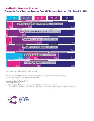
Classification • REAL/WHO Classification: • Nodular Lymphocyte Predominance • Calssical: • Lymphocyte-rich • Mixed cellularity • Nodular Sclerosis (50-80%) • Lymphocyte depletion (rare) • Not classifiable cases • Relation to Non Hodgkin’s Lymphomas • HL and Follicular lymphoma • CLL can progress to HL • Difficult distinction , diffuse large B-cell, anaplastic large cell.
Staging • Ann Arbor Staging system
Nodular Lymphocyte Predominance HL • 5% Of all HL • Children , young and middle aged • M:F 3:1 • Solitary enlarged peripheral LN, neck , groin and axilla • Mediastinal LN very rare • Relapse is common 19-32% • Median time for relapse 4 y • Stage I disease excellent 95% • Stage VI 24% • Treatment : ? Surgery for I, limited radiation.
Pathology • Nodal architecture is obliterated • Multiple, crowded, large, dark-staining nodules of small lymphocyte • Eosinophils and plasma cells scanty • Lympho-histiocytic ( L&H ) cells • CD20+, bcl-6+, and EMA+ • CD30- • CD3+ T-cells form rosettes around the L&H cells • PCR of microdissected L&H cells showed clonal rearrangement of Ig genes (B-cell nature)
Pathology • Trasformation to Large cell Lymphoma: • 3-4% transform to diffuse large cell lymphoma • Localized disease, favorable prognosis • Overgrowth of L&H cells or NHL • Diffuse Lymphocyte predominance HL: • Rare • Minor nodular component on reticulin stain • More likely to be stage III or IV • Difficult to differentiate with T cell rich B cell lymphoma and lymphocyte rich classical HL
Classical Hodgkin’s Lymphoma • Presents with lymphadenopathy • Extranodal disease is uncommon • Spread: • Contiguous pattern • Similar to carcinomas • To spleen following para aortic LN • Then blood borne spread • The neoplastic Reed-Sternberg cells • Diagnostic R-S cells • Mononuclear R-S cells • Lacunar cells • Pleomorphic R-S cells • Mummified cells
Reed-Sternberg Cells • Histogenesis controversial • Histiocyte, interdigitating dendritic cells, Follicular dendritic cells, myeloid cells and lymphocyte • B-cell lineage • B-cell specific activator protein and sometimes CD20 • Ig gene rearragement • Immuno: • CD45RB (LCA) – • CD30+, CD15+(para nuclear globules with or without membranous staining) • B-cell markers , like CD20 occasionally • Genetics: • Ig gene rearrangement in single neoplastic cell microdissection
Classical Hodgkin’s Lymphoma • Association with EBV: • 40-50% of cases in the West, and 100% in underdeveloped countries and patients with AIDS • MC type 60%, NS type 35% • Stronger association in the head and neck region and at the extremes of life
Nodular Sclerosis • 5-year survival 80% • Pathology: • The capsule is thickened with multiple fibrous bands and nodularity • Lacunar cells with small lymphocytes, plasma cells, eosinophils, neutrophils and histiocytes • Syncytial variant of NSHL • Necrosis, geographic • The cellular phase: lack fibrous bands • Subclassification/grading: • British National Lymphoma Investigation (BNLI) • Grade I (54% 5-year survival) • Grade II (37% 5-year survival ): • Any of : > 25% of cellular nodule contain numerous bizarre R-S cells • >25% of cellular nodules show lymphocyte depletion • >80% of cellular nodules show a fibrohistiocytic pattern
Mixed Cellularity • Cervical and supraclavicular LN • Complete remission in 70% • Pathology: • Background of small lymphocytes, plasma cells, eosinophils, and histiocytes • Mononuclear and diagnostic R-S cells • Focal necrosis, secondary fibrosis
Lymphocyte-rich HL • Often early stage disease • Uncommon in the mediastinum • Worse survival than N-LPHL • 5-year survival 80% • Pathology: • Mononuclear and rare diagnostic R-S cells • Not L&H cells
Lymphocyte depletion HL • Prognosis poor , 80% dying within 3 years • Pathology: • 2 recognized types • Diffuse fibrosis type- rare R-S cells • Reticular type- abundant diagnostic and pleomorphic R-S cells
Anaplastic Large Cell Lymphoma, Hodgkin’s-like • Young adult with LN involvement and large mediastinal mass • Patients do poorly with therapy for HL, but good with aggressive chemo for NHL • Pathology: • Cohesive growth of large bizarre neoplastic cells • Architecture resemble HL, capsular thickening , nodular growth, sclerosis band • Admixed reactive cells • Most called nodular sclerosis, syncytial, NS2 or LDHL , reticular type • Immuno: like ALCL • CD30+, LCA+/-, EMA+/-, CD15+/-, CD3-/+
Special morphologic features of classical HL • Interfollicular HL • Can be in any type. Inter follicular growth • Foamy Histiocytes in HL • Confusion with xanthogranulomas • Epithelioid histiocytes • Mimic the pattern of Lennert’s lymphoma • HL in monocytoid B-cell clusters • Treatment altered HL • In the Spleen: • The number of nodules should be counted • First affect the pulp, in a periarteriolar location or in the marginal zone • In the Liver: • First in the portal areas • In the marrow: • Focal or diffuse fibrosis with non specific infiltrates • Marrow eosinophilia
Prognostic Factors in Classical HL • Stage • Histologic type • Sex and age: male and old , worse prognosis • B symptoms • Total tumor burden • Response to treatment • Vascular invasion • Lack of CD15, expression of CD20