Download
1 / 7
70 likes | 182 Views
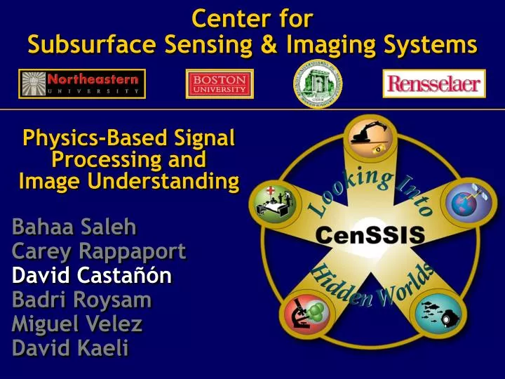
Center for Subsurface Sensing & Imaging Systems. Physics-Based Signal Processing and Image Understanding Bahaa Saleh Carey Rappaport David Castañón Badri Roysam Miguel Velez David Kaeli. Biological-Medical Applications. Environmental-Civil Applications. Bio-Med. Enviro-Civil. S2.

E N D
Center forSubsurface Sensing & Imaging Systems Physics-Based Signal Processing and Image Understanding Bahaa Saleh Carey Rappaport David Castañón Badri Roysam Miguel Velez David Kaeli
Biological-Medical Applications Environmental-Civil Applications Bio-Med Enviro-Civil S2 S3 S4 S1 S5 Breast Imaging Radiation therapy Vulnerable plaque Pollution monitoring Mine Detection Retinal surgery Coral Reef Monitoring Cellular Imaging Functional Imaging ValidatingTestBEDs S1 S3 S4 S2 S5 Application instantiations Algorithm requirements Common mathematical structures General information extraction approaches R1 R3 R2 in CenSSIS L3 L2 R2 FundamentalScience L1
1 2 .. I 1 2 .. J Medium Clutter object R2A. Multiview Tomography • Focus of research: Image formation from tomographic measurements • Modalities of interest in CenSSIS: • EIT, ERT, DOT, GPR, diffraction tomography, CT, ultrasound, elastography • Diverse applications approached through a common view: statistical physics-based inverse scattering • Inverse problem goals + physics model + solution representation + robust decision-directed algorithm • Objective: Improved resolution, signal-noise in subsurface imaging Y = T(, S, ) + w
R2A Highlights This Past Year • Continued refinement of techniques for transition to System Applications • EIT, DOT for breast imaging instruments in S3 • Multi-detector DCT for detection of coronary stenosis • Expansion of algorithm approaches • Nonlinear inversion approaches • Boundary element techniques for EIT • 4-D inverse problems (DOT, PET, others) • Multispectral DOT with spectral priors • Multisensor, multi-modal inversion • Time reversal high-resolution imaging • Inversion techniques for new modalities • Quantum OCT, Nano-imaging
Brief Overview of R2A Posters • Motivating Application: Breast Cancer • Student Posters • R2A p1 S. Laxminarayan et al, “Controlling Dimensionality in a Systems Approach to Dynamic Modeling” • R2A p2 E. Vidolova et al, “Hemodynamically Constrained Dynamic Diffuse Optical Tomography Under Mammographic Compression ” • R2A p5 G. Boverman et al, “Modeling and Electrode Contact Compensation for EIT in a Mammography Geometry” • R2A p6 C. Tamma, N. Liu et al, “ACT4: A High-Precision, Multi-frequency Electrical Impedance Tomograph” • R2A p7 T. Kao et al, “Regional Admittivity of Spectra with Tomosynthesis Image for Breast Cancer Detection” • R2A p8 R. Kulkarni et al, “A Layered Model for Breasts in Electrical Impedance Tomography”
R2A Posters Overview - 2 • Other Multi-View Tomography Posters • R2A p3 F. Gruber et al, “New Aspects in Electromagnetic Information Theory” • R2A p4 Z. Liang et al, “Image Enhancement in Detection of Coronary Stenosis by MDCT” • R2A p9 B. Alacam et al, “Reconstruction of Spatially Resolved Pharmacokinetic Rate Images of Optical Fluorophores from NIR Measurements ” • R2A p10 M. Guven et al, “Flourescence Optical Tomography with a Priori Information ” ConventionalImage of calcium and blood vessel Enhanced MDCT vessel
S3 S1 S5 S4 S2 Association with Systems Areas Cell Counting, Quadrature Tomography IMRT, Optical Biopsy, 4-D Inverse Problems Multi-view Tomography for EIT, DOT, Elastography Change Detection, Mulitspectral Classification Crosswell tomography, Rough Surface Imaging, Time Reversal Imaging