Download
1 / 152
1.57k likes | 1.62k Views
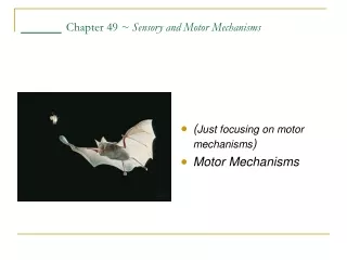
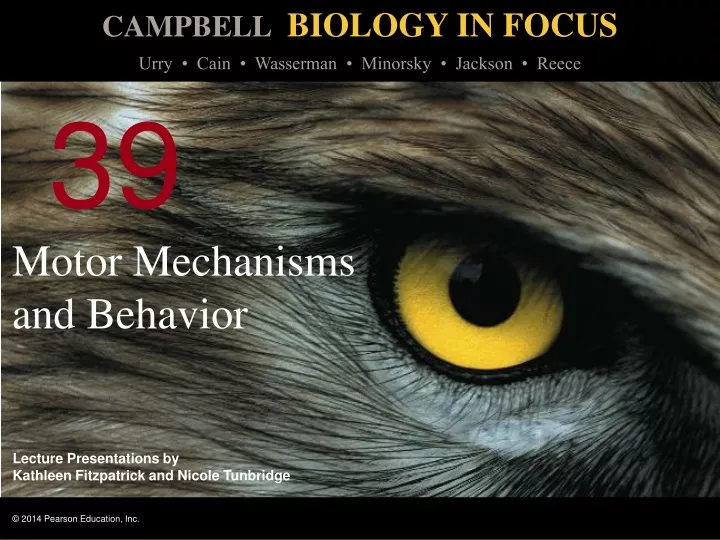
Motor Mechanisms and Behavior. 0. 39. Overview: The How and Why of Animal Activity. Fiddler crabs feed with their small claw and wave their large claw Why do male fiddler crabs engage in claw-waving behavior? Claw waving is used to repel other males and to attract females.

E N D
Overview: The How and Why of Animal Activity Fiddler crabs feed with their small claw and wave their large claw Why do male fiddler crabs engage in claw-waving behavior? Claw waving is used to repel other males and to attract females Video: Albatross Courtship Video: Boobies Courtship Video: Giraffe Courtship
A behavior is an action carried out by muscles under control of the nervous system Behavior is subject to natural selection
Concept 39.1: The physical interaction of protein filaments is required for muscle function Muscle activity is a response to input from the nervous system Muscle contraction is an active process; muscle relaxation is passive
Muscle cell contraction relies on the interaction between protein structures Thin filaments consist of two strands of actin coiled around one another Thick filaments are staggered arrays of myosin molecules
Vertebrate Skeletal Muscle Vertebrate skeletal muscle movesbones and the body andis characterized by a hierarchy of smaller and smaller units A skeletal muscle consists of a bundle of long fibers, each a single cell, running parallel to the length of the muscle Each muscle fiber is itself a bundle of smaller myofibrils, which contain thick and thin filaments
Skeletal muscle is also called striated muscle because the regular arrangement of myofibrils creates a pattern of light and dark bands The functional unit of a muscle is called a sarcomere and is bordered by Z lines
Figure 39.2 Muscle Bundle of muscle fibers Nuclei Single muscle fiber (cell) Plasma membrane Myofibril Z lines Sarcomere TEM Thick filaments (myosin) 0.5 m M line Thin filaments (actin) Z line Z line Sarcomere
Figure 39.2a Muscle Bundle of muscle fibers Nuclei Single muscle fiber (cell) Plasma membrane Myofibril Z lines Sarcomere
Figure 39.2b Sarcomere TEM Thick filaments (myosin) 0.5 m M line Thin filaments (actin) Z line Z line Sarcomere
Figure 39.2c Sarcomere TEM 0.5 m
The Sliding-Filament Mechanism of Muscle Contraction According to the sliding-filament model, filaments slide past each other longitudinally, causing an overlap between thin and thick filaments Video: Cardiac Muscle
Figure 39.3 Sarcomere Relaxed muscle Z M Z M Z Z Contracting muscle 0.5 m Fully contracted muscle Contracted sarcomere
Figure 39.3a Sarcomere M Z Z 0.5 m
Figure 39.3b Sarcomere M Z Z 0.5 m
Figure 39.3c 0.5 m Contracted sarcomere
The sliding of filaments relies on interaction between actin and myosin The “head” of a myosin molecule binds to an actin filament, forming a cross-bridge and pulling the thin filament toward the center of the sarcomere Muscle contraction requires repeated cycles of binding and release
Glycolysis and aerobic respiration generate the ATP needed to sustain muscle contraction Video: Myosin and Actin
Figure 39.4 1 5 2 3 P P P i i i 4 Thin filaments Thick filament Thin filament Myosin head (low energy configuration) ATP ATP Thick filament Myosin- binding sites Thin filament moves toward center of sarcomere. Actin ADP Myosin head (high energy configuration) Myosin head (low energy configuration) ADP ADP Cross-bridge
Figure 39.4a 1 Thin filament Myosin head (low energy configuration) ATP ATP Thick filament
Figure 39.4b 2 P i Myosin- binding sites Actin ADP Myosin head (high energy configuration)
Figure 39.4c 3 P i ADP Cross-bridge
Figure 39.4d 4 Thin filament moves toward center of sarcomere. Myosin head (low energy configuration)
The Role of Calcium and Regulatory Proteins Theregulatory protein tropomyosin and the troponin complex, a set of additional proteins, bind to actin strands on thin filaments when a muscle fiber is at rest This prevents actin and myosin from interacting
Figure 39.5 Ca2-binding sites Tropomyosin Actin Troponin complex (a) Myosin-binding sites blocked Ca2 Myosin- binding site (b) Myosin-binding sites exposed
For a muscle fiber to contract, myosin-binding sites must be uncovered This occurs when calcium ions (Ca2) bind to the troponin complex and expose the myosin-binding sites Contraction occurs when the concentration of Ca2 is high; muscle fiber contraction stops when the concentration of Ca2 is low
The stimulus leading to contraction of a muscle fiber is an action potential in a motor neuron that makes a synapse with the muscle fiber The synaptic terminal of the motor neuron releases the neurotransmitter acetylcholine Acetylcholine depolarizes the muscle, causing it to produce an action potential Animation: Muscle Contraction
Figure 39.6 1 2 3 4 7 6 5 Axon of motor neuron Synaptic terminal T tubule Sarcoplasmic reticulum (SR) Mitochondrion Myofibril Plasma membrane of muscle fiber Ca2 released from SR Sarcomere Synaptic terminal of motor neuron Plasma membrane Synaptic cleft T tubule Sarcoplasmic reticulum (SR) ACh Ca2 pump Ca2 ATP CYTOSOL Ca2
Figure 39.6a Synaptic terminal Axon of motor neuron T tubule Sarcoplasmic reticulum (SR) Mitochondrion Myofibril Plasma membrane of muscle fiber Ca2 released from SR Sarcomere
Figure 39.6b 1 2 3 4 7 6 5 Synaptic terminal of motor neuron Plasma membrane Synaptic cleft T tubule Sarcoplasmic reticulum (SR) ACh Ca2 pump Ca2 ATP CYTOSOL Ca2
Action potentials travel to the interior of the muscle fiber along transverse (T) tubules, infoldings of the plasma membrane The action potential along T tubules causes the sarcoplasmic reticulum (SR), a specialized endoplasmic reticulum, to release Ca2 The Ca2 binds to the troponin complex on the thin filaments, initiating muscle fiber contraction
When motor neuron input stops, the muscle cell relaxes Transport proteins in the SR pump Ca2 out of the cytosol Regulatory proteins bound to thin filaments shift back to the myosin-binding sites
Amyotrophic lateral sclerosis (ALS; also known as Lou Gehrig’s disease) interferes with the excitation of skeletal muscle fibers; this disease is usually fatal Myasthenia gravis is an autoimmune disease that attacks acetylcholine receptors on muscle fibers; treatments exist for this disease
Nervous Control of Muscle Tension Contraction of a whole muscle is graded, which means that the extent and strength of its contraction can be voluntarily altered There are two basic mechanisms by which the nervous system produces graded contractions Varying the number of fibers that contract Varying the rate at which fibers are stimulated
In vertebrates, each motor neuron may synapse with multiple muscle fibers, although each fiber is controlled by only one motor neuron A motor unit consists of a single motor neuron and all the muscle fibers it controls
Figure 39.7 Spinal cord Motor unit 2 Motor unit 1 Synaptic terminals Nerve Motor neuron cell body Motor neuron axon Muscle Muscle fibers Tendon
Recruitmentof multiple motor neurons results in stronger contractions A twitch results from a single action potential in a motor neuron More rapidly delivered action potentials produce a graded contraction by summation
Figure 39.8 Tetanus Summation of two twitches Tension Single twitch Time Action potential Series of action potentials at high frequency Pair of action potentials
Tetanus is a state of smooth and sustained contraction produced when motor neurons deliver a volley of action potentials
Types of Skeletal Muscle Fibers There are several distinct types of skeletal muscles, each of which is adapted to a particular set of functions They are classified by the source of ATP powering the muscle activity and the speed of muscle contraction
Oxidative and glycolytic fibers are differentiated by their energy source Oxidative fibers rely mostly on aerobic respiration to generate ATP These fibers have many mitochondria, a rich blood supply, and a large amount of myoglobin Myoglobinis a protein that binds oxygen more tightly than hemoglobin does
Glycolytic fibers use glycolysis as their primary source of ATP Glycolytic fibers have less myoglobin than oxidative fibers and tire more easily In poultry and fish, light meat is composed of glycolytic fibers, while dark meat is composed of oxidative fibers
Fast-twitch and slow-twitch fibers are differentiated by their speed of contraction Slow-twitch fibers contract more slowly but sustain longer contractions All slow-twitch fibers are oxidative Fast-twitch fibers contract more rapidly but sustain shorter contractions Fast-twitch fibers can be either glycolytic or oxidative
Most human skeletal muscles contain both slow-twitch and fast-twitch muscles in varying ratios Some vertebrates have muscles that twitch at rates much faster than human muscles In producing its characteristic mating call, the male toadfish can contract and relax certain muscles more than 200 times per second
Other Types of Muscle In addition to skeletal muscle, vertebrates have cardiac muscle and smooth muscle Cardiac muscle, found only in the heart, consists of striated cells electrically connected by intercalated disks Cardiac muscle can generate action potentials without neural input
In smooth muscle, found mainly in walls of hollow organs such as those of the digestive tract, contractions are relatively slow and may be initiated by the muscles themselves Contractions may also be caused by stimulation from neurons in the autonomic nervous system
Concept 39.2: Skeletal systems transform muscle contraction into locomotion Skeletal muscles are attached in antagonistic pairs, the actions of which are coordinated by the nervous system The skeleton provides a rigid structure to which muscles attach Skeletons function in support, protection, and movement