Download
1 / 48
490 likes | 593 Views
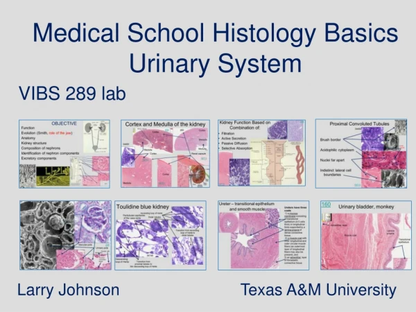
Medical School Histology Basics Introduction to Microscopy. VIBS 289 lab. Larry Johnson Texas A&M University. Objectives. Learn the difference in magnification and resolution Learn about different types of staining for LM and observe details of cells by EM

E N D
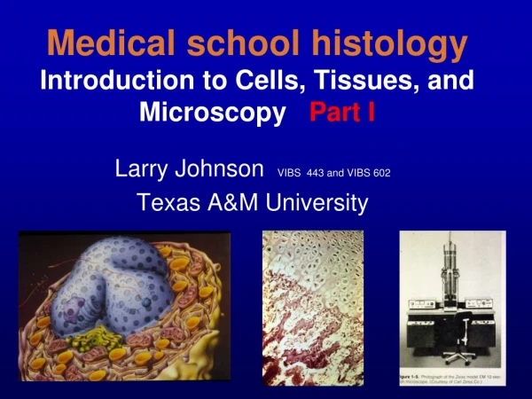
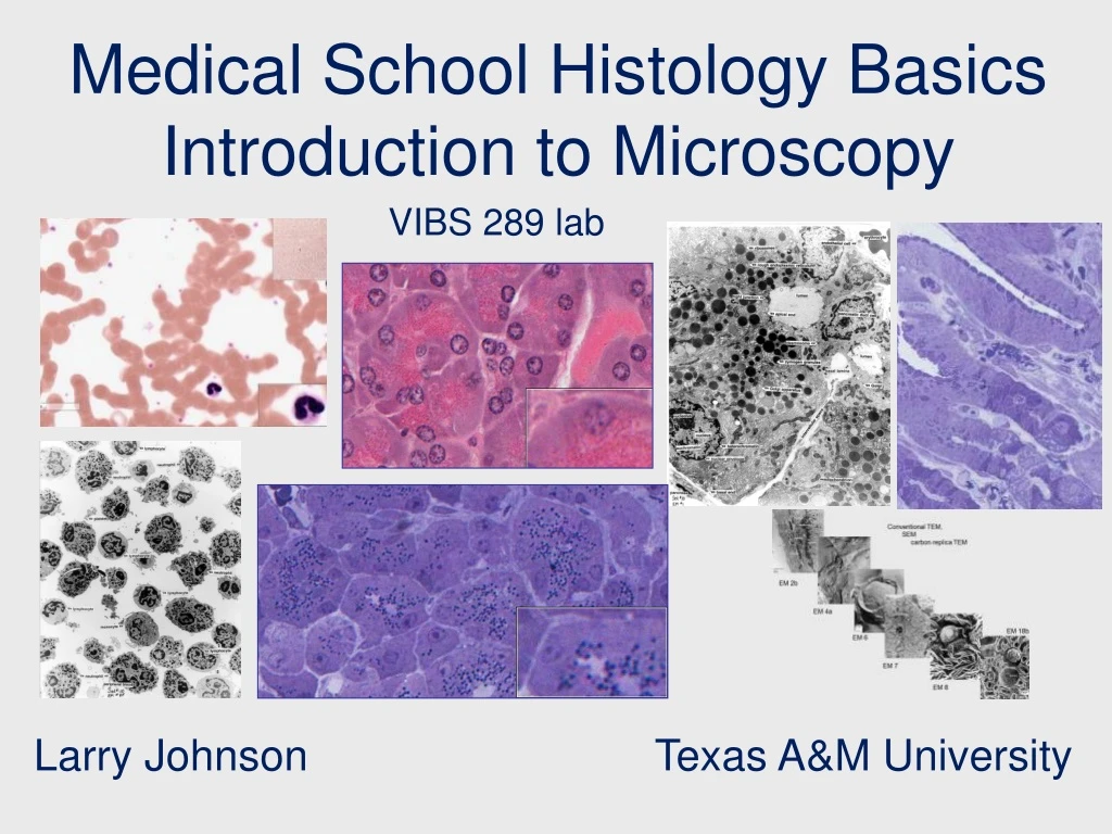
Medical School Histology Basics Introduction to Microscopy VIBS 289 lab Larry Johnson Texas A&M University
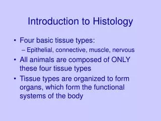
Objectives • Learn the difference in magnification and resolution • Learn about different types of staining for LM and observe details of cells by EM • Learn how cell/organelles look different at different magnifications • Learn about different types of EM
Use your atlas! Use your atlas! Use your atlas! Use your atlas! pancreas blood stomach testis
MAGNIFICATION VS. RESOLUTION • MAGNIFICATION - INCREASE IN IMAGE SIZE 2. RESOLUTION - SMALLEST DISTANCE BETWEEN TWO POINTS THAT CAN BE SEEN (DISTINGUISHED) RESOLUTION is CALCULATED BY 0.61 (WAVELENGTH)/NUMERICAL APERTURE 0.25 um FOR LIGHT MICROSCOPE 0.1 nm FOR ELECTRON MICROSCOPE
110 Peripheral blood smear (Leishman-Giemsa)basophil, and neutrophils
Slide 113 human blood Neutrophils
Slide Histo021 human blood Platelets Nucleus Neutrophils Red blood cells Neutrophil cytoplasm will merely have a granular appearance Neutrophils
EM 8f: Peripheral blood cells; 9,000x • Monocyte • Lymphocyte • Neutrophil Granules Neutrophils
Pancreas 158 In H&E staining, the acid dye is eosin (stains proteins red) and the basic dye is a completed form of hematoxylin (stains ribosomes and nuclei blue). Hence, color provides distinguishing characteristics. Islets of Langerhans = light-staining endocrine portion produces insulin Acinar cells = exocrine produces pancreatic enzymes
Pancreas 158 Islets of Langerhans Secretory granules are red as they are protein rich with enzymes Base cytoplasm is blue with ribosomes as in RER
Pancreas, monkey (toluidine blue) 156 The entire pancreatic acinar cell is blue with varying intensities depending on the density of structures. Shape, size, and darkness are used to identify structures. Secretory granules
158 Secretory granules Secretory granules are red as they are protein rich Base cytoplasm is blue with ribosomes as in RER 156 Smooth cytoplasm region = high density of ribosomes in this case
145 Fundic stomach Mucosa Connective tissue of submucosa
145 Fundic stomach: mucosa Chief cellsParietal cells Chief cells
244 Fundic stomach, rabbit (toluidine blue) Chief cellsParietal cells
244 Secretory granules in chief cells 145 Dark spots visible with toluidine blue staining are mitochondria in parietal cells . Nuclei Mitochondria are not distinguishable with H&E staining
19680 Toluidine blue staining Human testis - blood and lymph vessels
165 H & E staining and right insert toluidine blue staining – note differences in details of cytoplasm UT165 human testis Leydig cells 19680
EM 8f EM 12a EM 4c EM 6a Compare sizes of membranes ribosomes mitochondria as transmission electron microscopy (TEM) provides more cellular detail than light microscopy EM 2b EM 7
EM 8f: Peripheral blood cells; 9,000x • Monocyte • Lymphocyte • Neutrophil
EM 12a: Bone marrow; 13,200x. Note the reticular cell and developing red blood cells. • Reticular cell • Developing red blood cell
EM 4c: Intestinal absorption cell; 60,000x • Budding RER • Coated vesicle • Golgi • Mitochondria • Nucleus • Plasma membrane • Primary lysosome
EM 2b: Liver; 60,000x; cytoskeletal elements. Microtubes, microfilaments, and intermediate filaments can be compared in this cell, which has a high concentration of cortical microfilaments. • Microtube • Microfilaments • Intermediate filaments
EM 7: Ascites fluid; 80,000x. Clear examples of Golgi apparatuses with their cisternae and vesicles are present in this cell • Golgi apparatus • Ribosomes • Lipofuscin • Mitochondrion
EM 6a: Centriole-microtubules; 200,000x. Centriolar region of a cell showing both the stable, triplet microtubule arrays within the centriole, and the labile, individual microtubules originating from pericentriolar material. • Centriole • Stable microtubule • Labile microtubule
EM 4c 60,000x EM 8f 9,000x Compare sizes of membranes ribosomes mitochondria EM 12a 13,200x
EM 6a 200,000x EM 2b 60,000x Compare sizes of membranes ribosomes mitochondria EM 7 80,000x
Conventional TEM, SEM carbon replica TEM EM 2b EM 4a EM 18b EM 6 EM 7 EM 8
Conventional TEM, SEM carbon replica TEM EM 2b EM 4a EM 18b EM 6 EM 7 EM 8
Conventional TEM, SEM carbon replica TEM EM 2b EM 4a EM 18b EM 6 EM 7 EM 8
Conventional TEM, SEM carbon replica TEM EM 2b EM 4a EM 18b EM 6 EM 7 EM 8
In summary Use your atlas! Use your atlas! pancreas blood stomach testis
Questions Which microscope type/staining is/are better for observing cellular details: • Light microscopy/ H&E • Light microscopy/ toluidine blue • Transmission electron microscopy (TEM)/ typical EM staining • a and b • a, b, and c
Mexico USA Santa Elena Canyon Big Bend National Park, TX