Download
1 / 133
1.33k likes | 1.34k Views
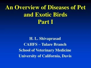
An Overview of Diseases of Pet and Exotic Birds Part II. H. L. Shivaprasad CAHFS – Tulare Branch School of Veterinary Medicine University of California, Davis. Psittacine Herpesviruses. Diverse group of viruses which infect a variety of psittacines – disease syndromes PsHV-1 and PsHV-2

E N D
An Overview of Diseases of Pet and Exotic BirdsPart II H. L. Shivaprasad CAHFS – Tulare Branch School of Veterinary Medicine University of California, Davis
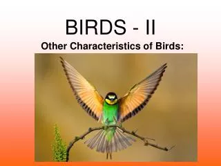
Psittacine Herpesviruses • Diverse group of viruses which infect a variety of psittacines – disease syndromes • PsHV-1 and PsHV-2 • Four genotypes and three serotypes • Diseases;Pacheco’s disease, Amazon tracheitis, Budgerigar herpesvirus (rare disease, “feather duster”) • Novel Herpesvirus; tracheitis, laryngitis, bronchitis, airsacculitis with syncytia formation and intranuclear inclusions – Bourke, Indian Ring-neck, others • Herpesvirus also associated with Papilloma’s
Psittacine Herpesviruses • Pacheco’s disease: acute viral disease of a variety of psittacines (common in 1980’s in US) • lesions: enlarged liver occasionally with petechiae, enlarged spleen, fluid filled intestine, diphtheretic membrane in oral cavity, esophagus, etc. • liver necrosis with or without inflammation, enteritis, stomatitis, esophagitis, pancreatitis, conjunctivitis, splenic and bursal necrosis, nephritis with intranuclear inclusion bodies • syncytia formation with inclusions in liver
Psittacine -Pacheco’s disease Hepatitis and splenitis
Psittacine -Pacheco’s disease Liver – Intranuclear inclusions
Other herpesviruses • Pigeon herpesvirus: • common in young squabs characterized by hepatitis, pancreatitis, esophagitis associated with intranuclear inclusions • conjunctivitis, enteritis, myocarditis, encephalitis, laryngitis, splenitis, etc. can also be seen • raptors and budgies susceptible • Finch herpesvirus • Duck Viral Enteritis (DVE) • Others : viruses of owls, falcons & eagles (same as pigeon herpes?), cranes, stork, starling, etc. • hepatitis associated with inclusion bodies
Finch Herpesvirus • Finch herpesvirus • disease of primarily of Gouldian finches • high mortality, conjunctivitis, tracheitis, bronchitis • cytomegalic cells and intranuclear inclusions • Herpesvirus demonstrated by EM and PCR • Novel virus (sequencing of DNA polymerase gene)
Herpesvirus infection in Finches • adults and both sexes affected • fibrinous exudate in conjunctiva, larynx, trachea, air sac, gray foci in lungs • increased pericardial fluid, enlarged liver and spleen, distended intestine, etc. • cytology of conjunctiva and trachea • karyomegalic cells with inclusions
Conjunctivitis, Gouldian Finches - Herpesvirus
Herpesvirus infection in Finches - Histopathology • proliferative conjunctivitis, sinusitis, rhinitis, laryngitis, tracheitis, bronchitis, bronchopneumonia, stomatitis and esophagitis • often with ulcers, fibrin exudation and bacteria • hypertrophied epithelial cells with intranuclear inclusion bodies
Conjunctivitis - G. Finch Herpesvirus inclusions
Severe tracheitis - G. Finch Herpesvirus inclusions
Psittacine Beak and Feather Disease • viral disease of many species of psittacines characterized by chronic feather and beak dystrophy • acute immunosuppression and sudden death in young birds due to secondary bacterial septicemia and fungal infections • etiology: circovirus • clin signs: dystrophic feathers first noticed of the powder down, progress to contour feathers, followed by primary, secondary tail and crest feathers, almost symmetrical
Psittacine Beak and Feather Disease - lesions • gross: abnormal and loss of feathers, sloughing of claws, beak necrosis, necrosis of oral mucosa, liver, bursa, thymus, etc. • micro: pterylitis and pulpitis associated with botryoid inclusions in macrophages, also in bursa, bone marrow, thymus, beak, claws, liver, pancreas, thyroid, testes, etc. • intranuclear inclusions in feather epithelium, intestine, esophagus, hepatocytes
PBFD – Cockatoos (Umbrella Cockatoo – normal). Note beak dystrophy in Moluccan
PBFD – Budgerigars and in a Love Bird
PBFD - Feather Botryoid inclusions (left) Intranuclear inclusions (arrows)
African Grey – Bursa of Fabricius PBFD botryoid inclusions
Other circoviruses • Pigeon circovirus • Pigeons/doves • Immunosuppression, feather dystrophy • Canary and Finch • Ducks, geese and swans • Gulls, starling, pheasant, ?
Circovirus infection in Pigeons • Disease in N. America, Europe and Australia • Probably worldwide? • Young birds most susceptible: 1- 4 months • Primarily immunosuppression and secondary infections • Lethargy, diarrhea, weight loss, sudden death, increased mortality in the flock, respiratory signs, poor racing performance • Bursal atrophy • some times with caseous exudate
Pigeon–Bursa of Fabricius. (Normal on top left). Circovirus infection(right two)
Pigeon – Bursa of Fabricius Circovirus botryoid inclusions
Circovirus infection in PigeonsCase Study (Duchatel et al., 2006) • Embryos, young, adults and breeders • Healthy adults (1 to 9 years): 65 % Pos. • Virus detected in ovary and testes and other organs • Embryos : 8/22 positive • Vertical transmission • Virus persists in respiratory system
Circovirus infection in PigeonsCase Study (Duchatel et al., 2006) • Testing of pharynx, feces and blood failed to detect all positive birds • Tissues collected at necropsy detected more positive birds • Young birds: • 15.8 % positive at 37 days of age • 100 % positive at 51 days of age • Acquired infection in the loft
Circovirus in Finches • 100 Gouldian Finches, mostly young • 20 adult and 12 young finches were added • 50 young and adult Gouldians died • Anorexia, sick nasal discharge, dyspnea • Bronchopneumonia, sinusitis, tracheitis • Lymphoid depletion in bursa, thymus • Inclusions in bursa of Fabricius, EM, ISH • Virus sequenced
Finch–Bursa of Fabricius. Circovirus infection(H&E and IHC)
Circoviruses phylogenic tree – Go-Goose, Du-Duck, BF-PBFD, Pi-Pigeon Gu-Gull, Ca-Canary, Fi-Finch
Papovavirus • Two genera are known to cause disease in psittacines and passerines • Papillomavirus and Polyomavirus • Papillomavirus: has been associated with cutaneous papillomas in wild finches (Fringilla) and an African Grey Parrot • Herpesvirus has been associated in psittacines with papillomas of cloaca • Conjunctiva, tongue, larynx, oral cavity, crop/esophagus, etc. ? • Polyomavirus: causes Budgerigar fledgling disease (BFD) in psittacines
Budgerigar Fledgling Disease • one of the most common diseases of psittacines (disease of 90’s,00’s?) and passerines • antibodies to BFDV been detected in chickens, but chickens are resistant to infection • etiology: polyomavirus, different strains such as psittacine, passerine, may exist • variety of psittacines, finches and canaries are susceptible • young psittacines are highly susceptible with very high mortality (30 - 100%) , also adults • feather dystrophy in budgerigars, acute death, digestive, neurological, respiratory, etc.
Budgerigar Fledgling Disease - lesions • gross: variation among psittacines and also passerines • in most of psittacines feather dystrophy, hemorrhages in skin, subcutis, skeletal muscle, heart, intestine, liver enlarged and mottled red or with white foci, splenomegaly, pale kidneys, ascites, lung congestion, pale carcass, etc. • in passerines, liver enlarged and mottled white, serosal or subserosal hemorrhage of intestine, pale myocardium, etc.
Budgerigar Fledgling Disease - lesions • micro: hemorrhages in various organs, necrosis in spleen, bursa, thymus and bone marrow, midzonal or random necrosis in liver, myocarditis, enteritis, nephritis, membranous glomerulopathy, pancreatitis, conjunctivitis, encephalomyelitis, ganglionitis (spinal), etc. • bluish karyomegalic inclusions in various tissues; epidermis, feather follicle epithelium, esophagus, kidney, macro/lympho of spleen, bursa, thymus, bone marrow, liver, etc. hepatocytes, myocytes, endothelial cells, glial cells, Purkinje cells, etc.
Polyomavirus infection Budgerigars
Psittacine – Feather epithelium Polyomavirus karyomegalic inclusions
Psittacine – Feather epithelium Polyomavirus karyomegalic inclusions
Psittacine – Feather pulp cavity Polyomavirus inclusions
Polyomavirus infection Hemorrhages: Cockatiel on top and a Parrot on right
Polyomavirus infection - Psittacine Hemorrhages: liver, intestine and spleen
Polyomavirus infection Hemorrhages: Heart and liver (Conure on left) & heart (Macaw top)
Polyomavirus infection - Psittacine Liver: hemorrhage & necrosis
Polyomavirus infection Spleen with inclusions (left) & epidermis (below)
West Nile Virus • Flavivirus (Family:Flaviviridae) • Corvids (Crow, raven, blue jay, magpie), grackle, house sparrow and finch, gulls, domestic geese, highly susceptible • Raptors; hawks, owls, eagles, falcon, osprey, kestrel • Psittacines; Rosella, lorikeet, cockatoo, conure, Australian parakeets, etc.
West Nile Virus and Birds Clinical signs • none, sudden death • anorexia, ruffled feathers, depression, weakness, lethargy • inability to walk or perch or stand or fly, droopy wings, do not respond to danger • neurological signs such as ataxia, torticollis, opisthotonus, tremors, circling, convulsions • loss of weight, anisocoria
West Nile Virus and Birds Gross Pathology • Depends on the species, none to marked • Hemorrhage in the calvarium, brain • Pale foci/streaks in the heart, hemorrhages • Hepatosplenomegaly, pale kidneys • Hemorrhage; esophagus, proventriculus, gizzard, intestine (diffuse ulcer), cloaca • pulmonary edema and congestion
WNV – geese, clinical signs Courtesy: Dr. Pokomonski
WNV – brain and spleen, crow Courtesy: Dr. Roberts
WNV – Gos Hawk. Heart and liver Courtesy: Dr. Dahlhausen
WNV in Psittacines • WNV not suspected by owners in 2004 (Calif.) • Clinical signs; most common - loss of weight, anorexia, lethargy and ruffled feathers • Sudden death in some, rarely neurological signs • Pathology: grossly none (hepatosplenomegaly, pale heart and kidneys?) • Microscopically myocarditis, nephritis, hepatitis, splenitis, enteritis, pancreatitis, encephalitis, etc. • Diagnosis: RT-PCR, Immunohistochemistry, Virus isolation, IHC, etc.
WNV– Rainbow Lorikeet Clinical signs (Courtesy: Dr. Luna)