Download
1 / 37
370 likes | 439 Views
Pathology of Rheumatic heart disease, infective endocarditis and valvular heart disease. Sufia Husain Department of Pathology KSU, Riyadh March 2016 Reference: Robbins & Cotran Pathology and Rubin’s Pathology. RHEUMATIC HEART DISEASE.

E N D
Pathology of Rheumatic heart disease, infective endocarditis and valvular heart disease Sufia Husain Department of Pathology KSU, Riyadh March 2016 Reference: Robbins & Cotran Pathology and Rubin’s Pathology
RHEUMATIC HEART DISEASE • Rheumatic heart disease is a heart disease caused by rheumatic fever. • Rheumatic heart disease can be • acute or • chronic
ACUTE Rheumatic Fever • Definition: Rheumatic fever (RF) is • an acute, immune mediated, multi-system inflammatory disease that occurs a few weeks after, group A-beta hemolytic streptococcal infection. • It is an acute post-streptococcal non-suppurative inflammatory disease with cardiac and extracardiac manifestations. • The inflammation is mainly in the heart, joints, central nervous system and skin. • Occurs in only 3% of patients with group A streptococcal pharyngitis. • It is seen mainly in children, 5 to 15 years of age. • Rheumatic fever is a major health problem in 3rd world countries and in crowded, low socioeconomic urban areas. • The incidence and mortality of rheumatic fever has declined over the past 30 years (due to improved socioeconomic condition and rapid diagnosis and treatment of strep. pharyngitis).
Etiopathogenesis: • The pathogenesis of RF remains unclear. • It is linked to streptococcal infection. Disease occurs 1 to 5 weeks after pharyngeal infection by Group A beta -Hemolytic Streptococcus. • It is most likely immune mediated in which the causative organisms (streptococci) result in the formation of antibodies which can cross react with certain antigens in the heart and joints (because they are similar to streptococcal antigen). Mechanism of its damage to the heart is not yet understood. • Repeated attacks or a single severe first attack can cause chronic rheumatic heart disease leading to congestive cardiac failure.
Cardiac manifestations of rheumatic FEVER also called as acute rheumatic heart disease or acute rheumatic carditis/ pancarditis There is a pancarditis in which there is inflammation in all three layers of the heart (endocardium, myocardium and pericardium) • Pericarditis: fibrinous or serofibrinous secretion in the pericardium. These secretion collect between the visceral and parietal pericardium like butter between two slices bread and therefore also called "bread and butter” pericarditis. • Myocarditis: many Aschoff bodies. Can cause sudden death. • Endocarditis: inflammation of the endocardium including the heart valves (valvulitis) and chordae tendineae. It results in fibrin deposition on valve leaflets forming tiny, pale thrombi along lines of closure called rheumatic vegetations. Mitral and aortic valve are mainly involved. Aschoff bodies/nodules are uncommon in the valves. This acute inflammation may either resolve completely or progress to scarring with development of chronic fibrotic deformities of the heart valves and chordae tendineae leading to chronic rheumatic heart disease many years later. • Subendocardial lesions can also be seen, commonly in the left atrium called as MacCallum plaques.
Aschoff bodies • The characteristic lesion of acute rheumatic fever are the Aschoff bodies. • Aschoff bodies are multiple tiny granulomatous lesions of the heart. They are situated next to small arteries and are characteristically seen in the myocardium (rheumatic myocarditis). • An Aschoff body, consists of: • a focus of eosinophilic collagen (representing the site of antibody-antigen reaction), • plump activated macrophages called Anitschkow/ caterpillar cells. Some of the macrophages become multinucleated to form Aschoff giant cells. • chronic inflammation. • Aschoff bodies are found mainly in the myocardium and pericardium. Uncommon in the endocardium and heart valves. • They ultimately "heal" by fibrosis resulting in a nodule of scar tissue.
Strep throat Antibody production Antibody cross-reaction with heart vegetations Aschoff body pericarditis
Rheumatic Vegetations: 1. Tiny (size of a pin’s head), sessile arranged in a row and firmly fixed with the underlying tissue.2. These are situated in the valve cusp, a few millimeters away from the free margin (this is the most traumatized area). tatic.wikidoc.org/1/1e/Rheumatic_fever_003.jpg
extra cardiac manifestations of Rheumatic Fever: Involvement of Other Organs • Joints: • Arthralgia • Migratory polyarthritis which is "fleeting arthritis" in the large joints e.g. knee, ankle, elbow wrist etc. It is self limiting with no chronic deformities. Aschoff bodies may be present in the synovial membrane, joint capsule, ligament etc. with joint effusion. • Skin: skin nodules, erythema marginatum. • Subcutaneous tissue: Rheumatic nodules mainly seen over the bony prominences e.g. knuckle, elbow, patella etc. • Neurologiocal disorder: Sydenhem’s chorea (St. Vitus' dance) characteristized by series of rapid involuntary purposeless movements of the face and arms. This occurs late in the disease. • Lung: uncommon, chronic interstitial inflammation and fibrinous pleuritis.
Acute Rheumatic Fever: Clinical features • Peak incidence: 5-15 years. • History of sore throat: symptoms start 10 days to 6 weeks after by group A Streptococcal pharyngitis • By that time the symptoms start the throat/ pharangeal cultures are usually negative. • Serum antistreptolysin O (ASO titer/antibodies to group A streptococcal antigens), anti-DNAase B, and antihyaluronidase are raised and provide evidence of a recent infection with group A Streptococcus. • Acute symptoms usually subside within 3 months • The mortality from acute rheumatic carditis is low. There is no specific test for rheumatic fever. The diagnosis is made based on the clinical findings. The clinical diagnosis is made when either • two major or • one major and two minor clinical features / criteria are met. This is called as the Jones criteria.
Acute Rheumatic Fever: Clinical Features continued…. JONES CRITERIA: Major criteria/ clinical features • Carditis (murmurs, pericardial friction rubs, weak heart sounds, tachycardia and arrhythmias cardiomegaly, pericarditis, and congestive heart failure) • Migratory polyarthritis of the large joints • Erythema marginatum of the skin • Subcutaneous nodules • Sydenhem’s chorea (St. Vitus' dance) Minor criteria / clinical features • Previous rheumatic fever • Arthralgia • Fever • Lab tests indicative of inflammatin — ESR (erythrocyte sedimentation rate), CRP (C-Reactive protein), leukocytosis • EKG changes
CHRONIC RHEUMATIC HEART DISEASE • The myocarditis and pericarditis components of RF typically resolve without permanent sequelae. • In contrast, the acute valvulitis or chordae tendinitis of rheumatic fever heals by fibrosis (scarring) and result in irreversible deformity of the involved cardiac valve and chordae tendineae. Severe valvular scarring develops months or years after acute RF. • Most harmful effect of rheumatic disease is due to involvement of cardiac valves. The valve leaflets develop diffuse fibrosis, become thickened, shrunken, and less movable which can lead to cardiac failure, thromboembolism and infective endocarditis.
CHRONIC RHEUMATIC HEART DISEASE Valves affected in chronic rheumatic heart disease: • Left side of heart is more commonly involved than the right. • mitral valve alone is most commonly affected • followed by combined mitral/aortic valve • Tricuspid valve is rarely affected. • Pulmonary valve is practically never affected. Type of damage • fibrosis of valve leaflets --> stenosis (Reduction of diameter) • fibrosis of chordae tendineae--> regurgitation (improper closure) • Therefore patient can have mitral stenosis (most common), mitral regurgitation, aortic stenosis and aortic regurgitation
Chronic Rheumatic heart disease: Clinical features • Manifests years or decades after the initial episode of rheumatic fever. • Signs and symptoms depend on the valve(s) involved: cardiac murmurs, hypertrophy, dilation, congestive heart failure, arrhythmia, thromboembolism and infective endocarditis. • Treatment may require valve surgery.
Complications of Chronic Rheumatic vavular Heart Disease • Bacterial infective endocarditis: the scarred valves of rheumatic heart disease provide an attractive environment for bacteria to grow. • Mural thrombi form in cardiac chambers. They give rise to thromboemboli, which can produce infarcts in various organs. • Congestive heart failure • Adhesive pericarditis • Atrial fibrillation.
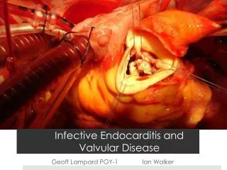
Infective Endocarditis (IE) • Definition: infection of the cardiac valves or mural surface of the endocardium, resulting in the formation of an adherent mass of thrombotic debris and micro-organisms. • Infective endocarditis is a particularly difficult infection to eradicate because of the avascular nature of the heart valves. • IE is Divided into: • Acute IE: • Is caused by highly virulent organisms (staphylococcus aureus) • infects even normal/healthy valves, • progresses rapidly, • Has little local host reaction. • Subacute IE: • It is an infection in a previously abnormal/ damaged valves by organisms of low virulence (-hemolytic streptococci viridans), • progresses slowly, • It induces a local inflammatory reaction. • Prognosis: depends to some extent on the offending organism and the stage at which the infection is treated. A third of cases of S. aureusendocarditis are still fatal.
Infective Endocarditis: Risk factors • In children: an underlying cardiac lesion (congenital heart disease is most common). • In adults: More than half of adults with bacterial endocarditis have no predisposing cardiac lesion. Mitral valve prolapse and congenital heart disease are the most frequent cause for bacterial endocarditis in adults. • Rheumatic heart disease. • Intravenous drug abusers can end up injecting micro-organisms intravenously when taking intravenous drugs, leading to IE. The tricuspid valve is infected in half of cases. About 50% of the IE in IV drug abusers are caused by S. aureus. • People with prosthetic valves are at high risk of developing IE . Prosthetic valve endocarditis is caused commonly by coagulase-negative staphylococci (e.g. S. epidermidis). • Transient bacteremia from any procedure may lead to infective endocarditis e.g. dental procedures, urinary catheterization, infected indwelling vascular catheters gastrointestinal endoscopy, and obstetric procedures. • The elderly (due to degeneration of heart valves e.g. calcific aortic stenosis), diabetics and pregnant women are at increased risk.
Infective Endocarditis: • Mitral valves are the most common sites of IE followed by aortic valve. • In IV drug users, the right side valves like the tricuspid are more commonly involved. • Vegetations may be single or multiple, involve one or more valve(s), differ in appearance according to the causative agent.
Infective Endocarditis: Clinical features • fever, fatigue, weight loss and chills. • Cardiac murmurs. • after 6 weeks: splenomegaly, petechiae, and clubbing of the fingers. • Positive blood culture for the organisms (only minority of cases remain negative). Complications: • valve ulceration and perforation, rupture of chordae tendineae, • arrhythmias, valvular regurgitation and congestive heart failure (due to destruction of a valve). • Septicemia or septic systemic embolization of infected vegetations which travel to multiple sites, causing infarcts or abscesses in many organs (e.g. neurologic deficits due to embolization to the brain or infarcts of the myocardium due to embolization to the coronary artery), • Pulmonary emboli is seen in tricuspid valve/ right sided endocarditis e.g. IV drug addicts. • Mycotic/infected aneurysms of vessels. • renal failure
D. Gross photograph illustrating healed endocarditis with perforations on bicuspid aortic valve Slide 13.43
Other types of endocarditis • Libman-Sacks endocarditis: Less common, noninfective, verrucous endocarditis associated with elevated levels of circulating immune complexes. Seen in patients with systemic lupus erythematosus • Endocarditis of carcinoid syndrome: Secretory products of carcinoid syndrome, especially 5-hydroxytryptamine can cause endocarditis. The endocardial plaques are seen in the right side of heart • Nonbacterial thrombotic endocarditis (marantic endocarditis)
Nonbacterial Thrombotic EndocarditisNBTE (marantic endocarditis) • Characterized by sterile vegetations (small masses of fibrin, platelets, and other blood components) on the leaflets of the cardiac valves. There is no infective organism. It is aseptic. • Pathogenesis/ association: • Subtle endothelial abnormalities. • Hypercoagulability. • Association with malignancy (50%) and other debilitating diseases. • Aortic valve most common site. The fibrin deposits are randomly arranged. • May embolize to different parts of the body including brain, but the emboli are sterile.
Diagrammatic comparison of the lesions in the four major forms of vegetative endocarditis. The rheumatic fever phase of RHD (rheumatic heart disease) is marked by a row of warty, small vegetations along the lines of closure of the valve leaflets. IE (infective endocarditis) is characterized by large, irregular masses on the valve cusps that can extend onto the cords. NBTE (nonbacterial thrombotic endocarditis) typically exhibits small, bland vegetations, usually attached at the line of closure. One or many may be present. LSE (Libman-Sacks endocarditis) has small or medium-sized vegetations on either or both sides of the valve leaflets. Robbin and Cotran Pathology
Valvular Heart Disease Two basic types: • Stenosis of valves: failure to open • Regurgitation of valves: Insufficiency or failure to close Both cause murmurs Causes • Most common cause of acquired valvular heart disease is post inflammatory scarring e.g. as a late result of rheumatic fever or secondary to various other inflammatory processes. • may be congenital. • can occur even with prosthetic cardiac valves • can be secondary to thrombus formation or infectious endocarditis.
Mitral valve 1. Mitral Valve Prolapse (picture) • is the most frequent valvular lesion in developed countries • seen in young women. • There is myxoid/mucoid degeneration of the valve which causes ballooning of mitral valves (floppy cusp), results in stretching of the mitral valve, producing a parachute deformity of the cusp with prolapse of the cusp into the atrium during systole. These changes produce a characteristic systolic murmur. • Pathogenesis unknown, can be a component of Marfan syndrome • Most patients asymptomatic but can result in mitral insufficiency and arrhythmias. • Patients are predisposed to infective endocarditis.
Mitral stenosis • Stenosis is more common than regurgitation. Mitral stenosis is most commonly due to rheumatic heart disease. • In mitral stenosis (picture): • Leaflets are thickened, fibrotic and fused leading to fish mouth/button hole deformity (stenosed valve looks like fish's mouth or button hole) • Increased pressure, dilatation and hypertrophy of left atrium. • secondary deposition of Ca++ • Pulmonary hypertension and lungs are firm and heavy (chronic passive congestion). • Right heart may be affected later (right ventricular hypertrophy).
mitral regurgitation • Is usually a result of rheumatic heart disease. • can also result from mitral valve prolapse, infective endocarditis, or damage to a papillary muscle from myocardial infarction etc • Leads to left vent. hypertrophy and dilatation.
Aortic stenosis Commonly caused by calcification and is called as calcific aortic stenosis.Calcific aortic stenosis can affect • Normal aortic valve as part of the aging degenerative process seen in persons older than 60 years of age • Congenital bicuspid aortic valve causing congenital aortic valvular disease • Valve affected and scarred by rheumatic heart disease http://keck.usc.edu/en/Education/Academic_Department_and_Divisions/Department_of_Medicine/Our_Divisions/Division_of_Cardiovascular_Medicine/Cardiovascular_Research_Unit/Clinical_Trials_and_Registries/Aortic_Stenosis.aspx
Aortic regurgitation/ insufficiency can be caused by: • Non-dissecting aortic aneurysm. • Rheumatic heart disease • Infective endocarditis • Syphilitic (luetic) aortitis (rare)
Right side of heart TRICUSPID VALVE • It is rarely involved in rheumatic heart disease along with the mitral and aortic valves. PULMONARY VALVE • Seen in congenital malformations e.g. tetralogy of Fallot. • Is very rarely involved in rheumatic heart disease